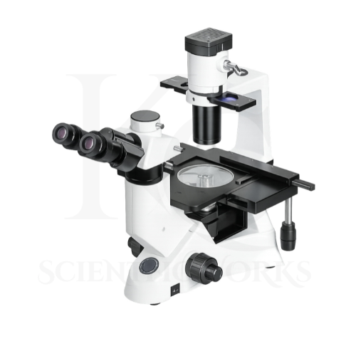
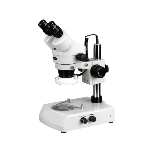
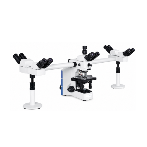
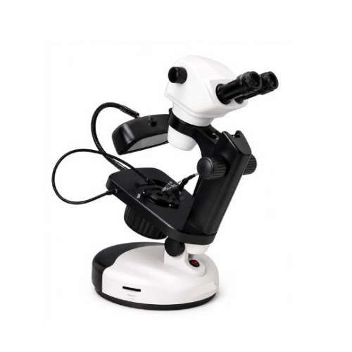
Biological Research Laboratories
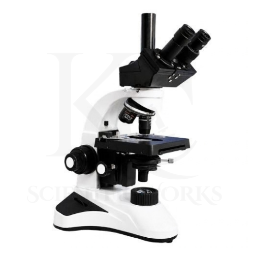
The Co-Axial Trinocular Microscope KCX-141 is suitable for routine biological specimen examination in scientific and educational laboratories.
Its compound optical system supports observation of compatible prepared slides.
College and University Laboratories
Colleges and universities can use the microscope for biology practical sessions, scientific demonstrations and laboratory training.
Digital Microscope Imaging
The trinocular camera port can support integration with compatible digital microscope cameras and adapters.
This enables specimen image capture and display when appropriate imaging equipment is installed.
Microbiology Education
The microscope can be used for observing appropriately prepared microbial specimens within the capabilities of its installed optical system.
Botany Laboratories
Botany departments can use the KCX-141 for examination of prepared plant tissue sections and other botanical specimens.
Zoology Laboratories
The microscope supports educational observation of suitable zoological specimens and prepared tissue slides.
Scientific Research Centers
Research centers can use the microscope for routine optical examination and documentation of compatible specimens.
Medical Laboratory Training
The KCX-141 can support educational observation of prepared biological slides and training in laboratory microscopy techniques.
Clinical diagnostic use would require appropriate product validation and applicable regulatory compliance.
Biology Practical Classes
Students can use the microscope to learn slide positioning, objective selection, focusing and specimen observation.
Scientific Demonstrations
Teachers and instructors can use the KCX-141 for laboratory demonstrations involving compound magnification and trinocular microscope operation.
Specimen Documentation
With compatible digital imaging accessories, the microscope can support photographic documentation of suitable laboratory specimens.
Laboratory Training Centers
Scientific training institutions can use the microscope to demonstrate binocular observation, objective selection and digital microscopy principles.
Working Principle
The Co-Axial Trinocular Microscope KCX-141 operates using the principles of compound optical magnification and transmitted-light microscopy.
A prepared specimen is positioned on the mechanical stage beneath the selected objective lens.
Light from the base-integrated illumination system passes through the condenser and specimen.
The selected objective lens produces a magnified optical image that is viewed through the binocular eyepieces.
The revolving nosepiece allows users to change objectives according to the required magnification.
The coaxial coarse and fine focusing controls adjust the relative position of the specimen and objective system to produce a sharp image.
The mechanical stage enables controlled movement of the specimen during examination.
The trinocular head includes a separate camera port through which a compatible imaging system may receive the microscope image.
Camera operation depends on the microscope's internal light-path arrangement and the selected imaging accessories.
Optical Magnification
Total optical magnification is calculated as:
Total Magnification = Eyepiece Magnification × Objective Magnification
The following table shows typical compound microscope combinations if 10X eyepieces and the listed objectives are installed.
| Objective Lens | Eyepiece | Total Magnification |
|---|
| 4X | 10X | 40X |
| 10X | 10X | 100X |
| 40X | 10X | 400X |
| 100X | 10X | 1000X |
These are illustrative magnification combinations, not verified KCX-141 specifications.
Microscope Compatibility and Usage
The KCX-141 Co-Axial Trinocular Microscope is designed for observation of compatible prepared biological slides and laboratory specimens.
It can be used in schools, colleges, universities, research laboratories and scientific training institutions.
For routine observation, users should position the prepared slide on the mechanical stage, select a suitable objective and adjust the coarse focusing control.
The fine focusing control can then be used to improve image sharpness.
For digital microscopy, a compatible camera and appropriate optical adapter can be connected to the trinocular port.
The microscope should be operated according to the final supplied technical configuration and product instructions.