Medical College Teaching
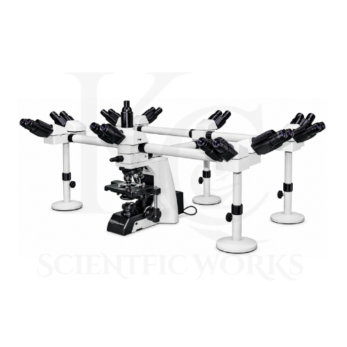
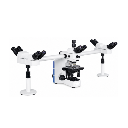
The Deca Head Microscope KCX-175 is suitable for medical colleges requiring shared microscope observation during educational laboratory sessions.
Its ten-person viewing arrangement allows instructors and students to examine the same specimen.
University Research Laboratories
Universities can use the KCX-175 for collaborative specimen examination, scientific demonstrations and laboratory teaching.
Pathology Training
The microscope can support educational observation of prepared histological and pathological teaching slides.
Any use for clinical diagnosis requires appropriate product validation and applicable regulatory compliance.
Histology Education
The microscope is useful for demonstrating prepared tissue sections during histology teaching and laboratory training.
Microbiology Education
The KCX-175 can support observation of appropriately prepared and stained microbial specimens during educational sessions.
Biological Research Laboratories
Biological research laboratories can use the microscope for collaborative examination of compatible prepared specimens.
Medical Laboratory Training
The microscope is suitable for teaching basic and advanced optical microscopy techniques in medical and laboratory training environments.
Collaborative Scientific Research
Research groups can use the multi-viewing system to discuss specimen features and compare observations during shared microscopy sessions.
Classroom Demonstrations
Instructors can use the KCX-175 to demonstrate microscopic structures to multiple students simultaneously.
Life Science Laboratories
The microscope can support educational and research examination of suitable prepared biological specimens.
Pharmaceutical Training
Pharmaceutical education and laboratory training programs can use the microscope for appropriate microscopy demonstrations and specimen examination.
Group Microscopy Sessions
The ten-person observation system is useful for workshops, laboratory demonstrations and group scientific discussions.
Scientific Training Centers
Scientific training institutions can use the KCX-175 to teach microscope operation, specimen focusing, objective selection and collaborative observation techniques.
Laboratory Specimen Demonstrations
The microscope supports group examination of prepared slides during laboratory instruction and research presentations.
Working Principle
The Deca Head Microscope KCX-175 operates using the principles of compound optical magnification, transmitted-light microscopy and multi-viewing optical distribution.
A prepared specimen is positioned on the double-layer mechanical stage beneath the selected objective lens.
Light from the 12V / 100W halogen illumination system passes through the condenser and specimen.
The Köhler illumination arrangement helps control the illumination field and supports even specimen lighting.
The selected infinity plan achromatic objective lens collects light from the specimen and forms an image through the microscope's infinity-corrected optical system.
The magnified image is then distributed through the multi-viewing optical arrangement to the main observation head and discussion heads.
This allows multiple users to examine the same microscopic field simultaneously.
The coaxial coarse and fine focusing controls adjust image sharpness, while the mechanical stage allows controlled specimen positioning.
The LED cursor provides a visible reference that can be used to indicate specific regions of the observed specimen during group discussions.
Optical Magnification Combinations
| Objective Lens | Eyepiece | Total Magnification |
|---|
| Infinity Plan 4X | WF10X | 40X |
| Infinity Plan 10X | WF10X | 100X |
| Infinity Plan 40X | WF10X | 400X |
| Infinity Plan 100X Oil | WF10X | 1000X |
Microscope Compatibility and Usage
The KCX-175 Deca Head Microscope is designed for observation of compatible prepared biological slides and laboratory specimens.
It can be used in medical colleges, universities, research institutions, educational laboratories and scientific training centers.
For routine observation, users should place a prepared specimen on the mechanical stage, select the appropriate objective lens and adjust the illumination.
The main operator can use the coaxial focusing controls to obtain a sharp image.
The multi-viewing system distributes the specimen image to the connected observation heads, allowing participants to observe the same field.
The LED cursor may be used to highlight selected specimen features during teaching and discussions.
The 100X oil immersion objective should be used with suitable immersion oil according to the product operating instructions.
The microscope should be operated according to its final supplied technical configuration and electrical requirements.