Advanced Endodontics
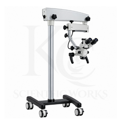
The Dental Microscope KCX-211 is intended to support magnified visualization during endodontic treatment.
Its optical system can help trained dental professionals examine the pulp chamber, locate small anatomical features and inspect the treatment field.
Root Canal Treatment
The microscope can support visualization of root canal openings, chamber anatomy and suitable treatment instruments during endodontic procedures.
Endodontic Retreatment
Magnified observation can assist trained clinicians when examining previously treated root canal systems and identifying visible obstructions or restoration materials.
Restorative Dentistry
The KCX-211 can support detailed visual examination during suitable restorative procedures.
Its magnification system allows clinicians to inspect restoration margins, tooth surfaces and prepared areas.
Aesthetic Dental Restorations
The microscope can assist with visualization during procedures involving veneers, inlays and other suitable restorations.
Dental Implantology
The microscope can provide magnified visualization during selected implant-related examinations and suitable soft-tissue procedures.
Dental Microsurgery
The KCX-211 can support magnified observation during suitable dental microsurgical procedures performed by appropriately trained clinicians.
Tooth Fracture Examination
The microscope may help clinicians visualize fine surface features and suspected fracture lines, subject to optical visibility and appropriate diagnostic assessment.
Dental Caries Examination
Magnification can support visual inspection of tooth surfaces and prepared cavities as part of a broader clinical examination.
Dental Education
Dental colleges can use the microscope for teaching magnified dental visualization, instrument handling and procedural observation.
Clinical Documentation
With compatible imaging accessories, the microscope can support photographic and video documentation of selected dental procedures.
Dental Research and Training
The microscope is suitable for appropriate dental research demonstrations, training activities and laboratory-based dental education.
Working Principle
The Dental Microscope KCX-211 operates using the principles of optical magnification, stereoscopic observation and coaxial illumination.
Light from the selected LED or halogen illumination system is directed toward the dental treatment area through the microscope's illumination arrangement.
The objective lens collects reflected light from the observed structures and forms an optical image.
The microscope's five-step Galileo magnification changer allows the operator to select different magnification levels by changing the internal optical configuration.
The binocular viewing system presents slightly different perspectives to the observer's eyes, providing stereoscopic depth perception.
The articulated arm enables the microscope to be positioned over the treatment area, while the fine-focus mechanism helps obtain a sharp image.
The adjustable illumination control allows the operator to modify brightness according to the observed surfaces and clinical requirements.
When an orange composite filter is selected, the illumination spectrum is modified to reduce blue light that may initiate polymerization in compatible light-curable dental composites.
For digital documentation, a compatible beam splitter can direct part of the optical image toward a camera system, depending on the installed accessories.
Magnification System
| Parameter | Specification |
|---|
| Magnification Type | Galileo-Type |
| Magnification Selection | Five Discrete Steps |
| Observation Method | Binocular Stereoscopic |
| Eyepieces | Wide-Field High-Eyepoint |
| Objective Working Distance | 200–400 mm Options |
| Actual Total Magnification | Depends on Eyepiece, Objective and Magnification Changer Configuration |
The five individual magnification values should be confirmed from the final KCX-211 optical configuration.
Dental Microscope Compatibility and Usage
The KCX-211 Dental Microscope is intended for use by appropriately trained dental professionals in compatible clinical and educational environments.
It can be used in dental clinics, dental colleges, dental hospitals, specialist treatment centers and training institutions.
Before use, the microscope should be positioned securely and adjusted to provide a suitable working distance and viewing angle.
The operator can select the required magnification and adjust illumination intensity according to the treatment area.
The orange filter may be selected during appropriate light-curable composite procedures.
Compatible imaging accessories may be installed when clinical documentation or educational display is required.
The microscope's actual clinical suitability depends on its final configuration, safety documentation, validation and applicable medical-device requirements.