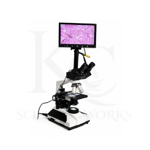
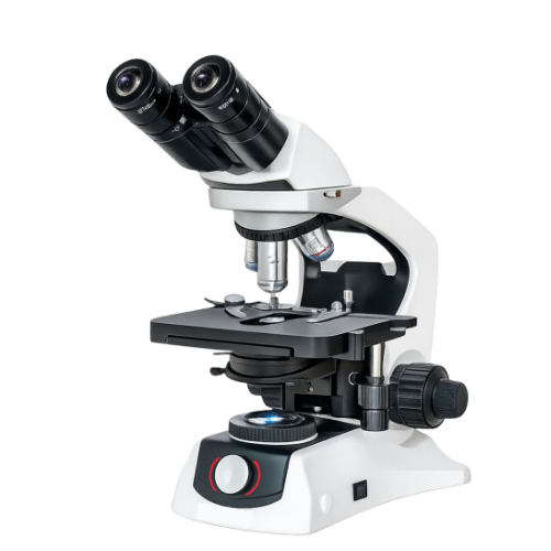
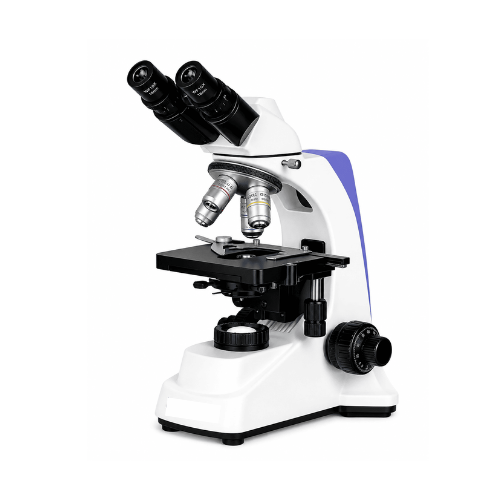
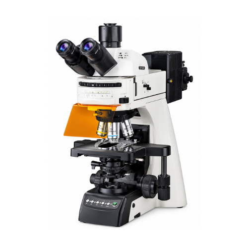
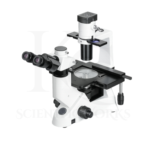
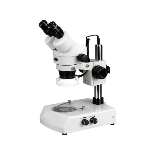
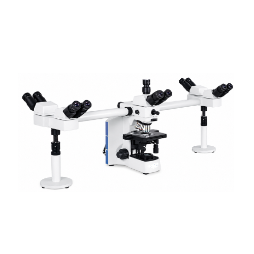
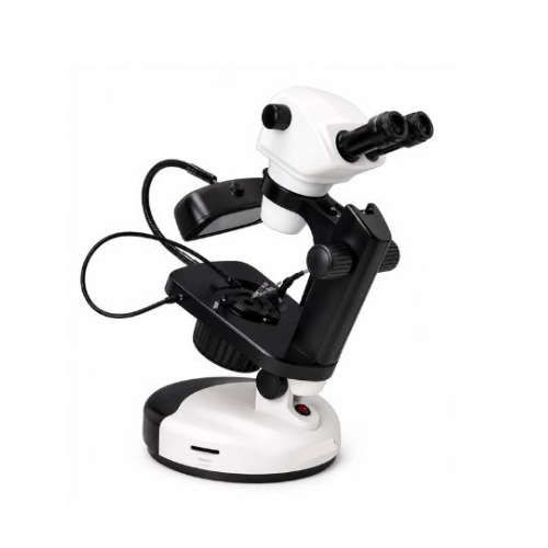
The Digital Microscope KCX-146 is designed to magnify biological specimens and present their images through both conventional optical eyepieces and an integrated digital display.
The microscope allows users to examine suitable cells, prepared tissues, microorganisms and other microscopic specimens through its plan achromatic optical system.
The 3 MP digital imaging system converts the microscope image into a digital signal that can be displayed on the 7-inch TFT LED screen.
This arrangement makes the microscope useful for scientific demonstrations, biology education, laboratory training and collaborative observation.
The integrated display allows instructors and students to examine the specimen image without requiring every observer to look through the eyepieces individually.
The microscope's mechanical stage supports controlled specimen positioning, while its coaxial focusing controls enable users to adjust image sharpness.
The LED or halogen illumination arrangement provides transmitted light for examining compatible prepared biological specimens.
The KCX-146 is therefore suitable for laboratories requiring a combination of optical microscopy and convenient digital visualization.
Applications
1. School Biology Laboratories
The KCX-146 Digital Microscope is suitable for school biology practical sessions and educational demonstrations.
Students can learn microscope operation, specimen focusing and biological observation using the eyepieces and integrated digital display.
2. College Laboratories
Colleges can use the microscope for routine biology practical classes, specimen examination and laboratory training.
3. University Research Laboratories
University laboratories can use the KCX-146 for routine optical examination and digital viewing of compatible prepared specimens.
4. Microbiology Education
The microscope supports educational observation of appropriately prepared and stained microbial specimens.
Its higher-magnification objectives can be used according to the optical requirements of the specimen.
5. Botany Laboratories
Botany departments can use the microscope for examining prepared plant tissue sections, cells and other botanical specimens.
6. Zoology Laboratories
The microscope can support observation of suitable prepared zoological specimens and tissue sections.
7. Histology Education
The KCX-146 can be used to demonstrate prepared tissue sections during histology teaching and laboratory training.
8. Medical Laboratory Training
The microscope is suitable for educational observation of prepared biological slides and training in basic microscopy techniques.
Clinical diagnostic use requires appropriate validation and applicable regulatory compliance.
9. Scientific Research Centers
Research laboratories can use the microscope for suitable biological specimen examination and digital observation.
10. Digital Microscopy Demonstrations
The integrated 7-inch display allows instructors to demonstrate microscopic structures to students and laboratory trainees.
11. Laboratory Specimen Documentation
The digital imaging system can support documentation of suitable microscopic specimens when the installed imaging functions permit image capture or recording.
12. Scientific Training Centers
Training institutions can use the microscope to demonstrate optical magnification, digital imaging, objective selection and specimen focusing.
13. Biological Cell Observation
The microscope can be used for examining appropriately prepared cells and selected cellular structures using conventional transmitted-light microscopy.
14. Laboratory Teaching and Group Observation
The integrated screen allows multiple people positioned near the microscope to view the same digital image during demonstrations.
Working Principle
The Digital Microscope KCX-146 operates using the principles of compound optical magnification, transmitted-light microscopy and digital image conversion.
A prepared biological specimen is positioned on the double-layer mechanical stage beneath the selected objective lens.
Light from the LED or halogen illumination system passes through the condenser and specimen.
The selected plan achromatic objective collects light from the specimen and forms a magnified optical image.
The image is viewed through the WF10X eyepieces using the binocular observation system.
For digital viewing, the microscope's imaging optics direct an image toward the 3 MP digital camera sensor.
The camera converts the optical image into electronic image data, which is processed and displayed on the integrated 7-inch TFT LED screen.
The operator can adjust the coarse and fine focusing controls to obtain a sharp image.
The revolving nosepiece allows users to change objective magnification according to the observation requirement.
The mechanical stage enables controlled movement of the specimen, allowing different regions of the prepared slide to be examined.
The integrated display provides real-time digital visualization for laboratory teaching and specimen observation.
Optical Magnification Combinations
| Objective Lens | Eyepiece | Total Optical Magnification |
|---|
| Plan Achromatic 4X | WF10X | 40X |
| Plan Achromatic 10X | WF10X | 100X |
| Plan Achromatic 40X | WF10X | 400X |
| Plan Achromatic 100X | WF10X | 1000X |
Note: The magnification values above refer to optical magnification through the eyepieces. Magnification on the digital screen also depends on the imaging optics, sensor and display configuration.
Digital Imaging System
| Parameter | Specification |
|---|
| Imaging Technology | Digital Video Imaging |
| Camera Resolution | 3 MP |
| Display Size | 7 Inches |
| Display Technology | TFT LED |
| Observation | Real-Time Digital Viewing |
| Optical Integration | Microscope Digital Imaging System |
| External Computer | Not Required for Routine Live Viewing |
| Image Capture / Storage | Depends on Installed Recording and Storage Functions |
| Power Supply | DC 12V |
| Backup Facility | Battery Backup Supported |
Microscope Compatibility and Usage
The Digital Microscope KCX-146 is designed for observation of compatible prepared biological slides and laboratory specimens.
It can be used in schools, colleges, universities, research laboratories and scientific training institutions.
For routine observation, users should place a prepared microscope slide on the mechanical stage and select a suitable low-power objective.
The coarse focusing control should be used to locate the specimen, followed by fine focusing for improved image sharpness.
The integrated digital display can be used for live viewing and educational demonstrations.
Higher magnification objectives may be selected according to the specimen and observation requirements.
For digital documentation, users should follow the operating instructions for the installed camera, storage and recording system.
The microscope should be operated using the specified DC 12V power arrangement and compatible accessories.