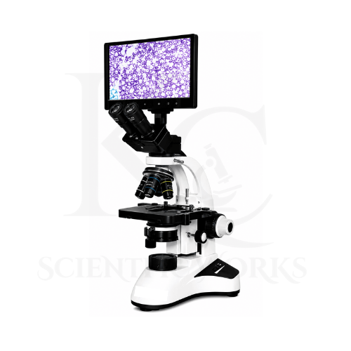
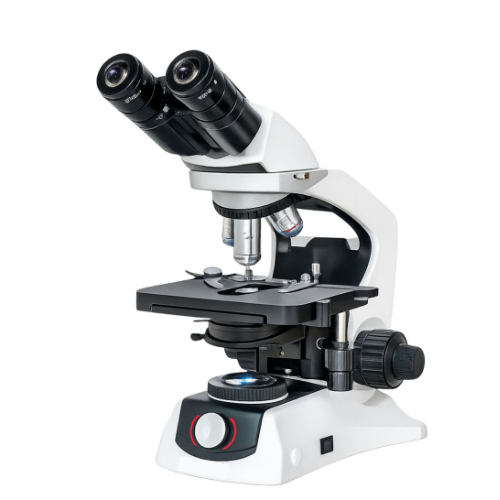
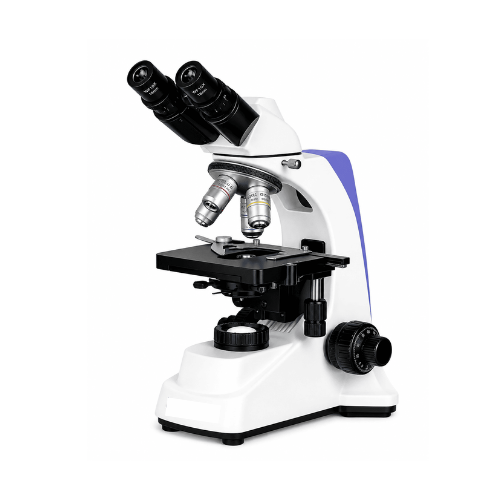
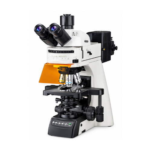
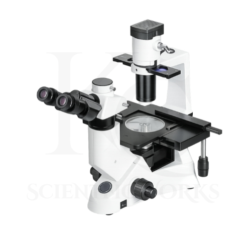
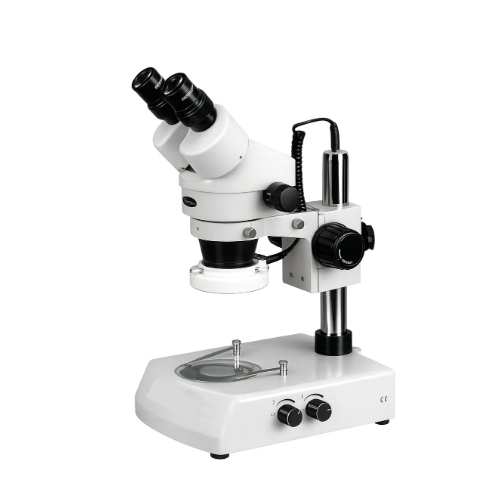
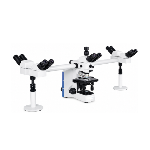
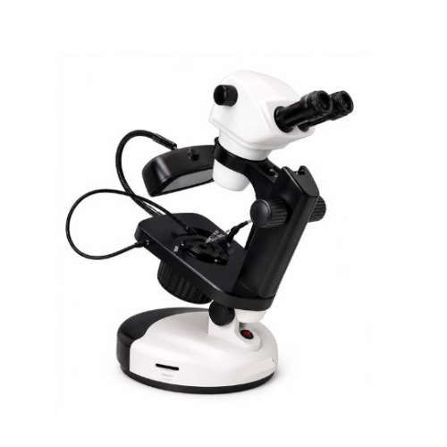
The Digital Video Microscope KCX-145 is designed to magnify compatible specimens and display their images directly on an integrated digital screen.
Unlike conventional microscopes that primarily rely on eyepiece observation, this microscope uses a built-in digital camera to present the specimen image on a 9.5-inch HD LED display.
The microscope's optical system enlarges the specimen image, while the digital camera converts the optical information into electronic image data.
The integrated display presents the resulting image in real time, allowing users to observe microscopic details during educational experiments, laboratory training and scientific demonstrations.
The adjustable LED illumination system provides light for specimen observation, while the coaxial focusing mechanism enables users to adjust image sharpness.
The mechanical stage supports controlled specimen positioning, allowing different sample regions to be examined.
The microscope's image capture capability can support laboratory documentation and educational reference.
The KCX-145 is therefore useful for laboratories and educational institutions requiring convenient digital viewing, practical specimen examination and collaborative observation.
Applications
1. School Biology Laboratories
The Digital Video Microscope KCX-145 is suitable for school biology practical classes and introductory microscopy demonstrations.
Students can observe suitable prepared specimens directly on the integrated digital screen.
2. College Laboratories
Colleges can use the microscope for scientific practical sessions, specimen observation and laboratory training.
3. University Laboratories
Universities can use the KCX-145 for suitable digital microscopy demonstrations and routine specimen examination.
4. Biological Specimen Observation
The microscope can be used to examine compatible prepared biological specimens within the capabilities of its optical system.
5. Microbiology Education
The microscope may support observation of appropriately prepared microbial specimens where the installed magnification and optical resolution are sufficient.
6. Botany Laboratories
Botany departments can use the microscope for examining suitable prepared plant specimens and selected microscopic structures.
7. Zoology Laboratories
The microscope can support observation of prepared zoological specimens and biological samples during educational activities.
8. Scientific Research Laboratories
Research laboratories can use the KCX-145 for routine digital examination of compatible specimens.
9. Laboratory Training Centers
Scientific training institutions can use the microscope to demonstrate digital imaging, optical focusing and specimen positioning.
10. Classroom Demonstrations
The integrated display allows instructors to demonstrate microscopic structures to students without requiring each observer to use an eyepiece.
11. Digital Specimen Documentation
The microscope's image capture capability supports documentation of suitable microscopic specimens for laboratory records and educational materials.
12. Material Observation
The KCX-145 may be used to examine compatible material samples and fine surface features where its optical arrangement, illumination and stage accommodate the specimen.
13. Electronics Education
The microscope may support educational examination of compatible small electronic components and circuit features, subject to the available working distance and illumination arrangement.
14. Laboratory Quality-Control Training
The microscope can support demonstrations of visual inspection and digital documentation techniques in suitable laboratory training environments.
15. Scientific Demonstrations
Teachers and laboratory personnel can use the microscope for explaining optical magnification, digital image formation and specimen examination.
16. Collaborative Laboratory Observation
The integrated screen enables nearby students and laboratory personnel to observe the same specimen image during group discussions.
Working Principle
The Digital Video Microscope KCX-145 operates using the principles of optical magnification, digital image sensing and real-time electronic display.
A compatible specimen is placed on the mechanical stage and positioned within the microscope's optical field.
Light from the adjustable LED illumination system illuminates the specimen.
The optical system collects light from the specimen and produces a magnified image.
The built-in megapixel camera sensor converts the optical image into electronic image data.
The imaging electronics process this information and send the resulting image to the integrated 9.5-inch HD LED display.
The user can observe the magnified specimen in real time on the screen.
The coaxial coarse and fine focusing controls allow the operator to adjust image sharpness.
The mechanical stage supports controlled specimen movement, enabling observation of different sample regions.
The image capture system allows selected specimen images to be recorded when the relevant storage and recording functions are available.
Digital Image Formation Process
Specimen Illumination: Adjustable LED lighting illuminates the sample.
Optical Magnification: The semi-plan optical system enlarges the specimen image.
Digital Image Conversion: The built-in megapixel camera converts the optical image into electronic data.
Image Processing: The digital imaging electronics process the captured information.
Live Display: The image appears on the integrated 9.5-inch HD LED screen.
Focusing and Positioning: Coaxial focusing and the mechanical stage allow users to refine the observed image.
Image Capture: Supported recording functions allow selected images to be documented.
Digital Imaging System
| Parameter | Specification |
|---|
| Digital Imaging Type | Integrated Digital Video Microscope |
| Camera | Built-in Megapixel Camera |
| Camera Resolution | Exact Megapixel Count Not Specified |
| Display Size | 9.5 Inches |
| Display Type | HD LED |
| Live Viewing | Supported |
| Specified Magnification | 100X |
| Image Capture | Supported |
| Video Recording | Not Specified |
| Storage Capacity | Not Specified |
| USB / HDMI Connectivity | Not Specified |
| External Computer | Not Required for Basic Live Viewing |
| Optical System | Semi-Plan |
| Illumination | Adjustable LED |
| Focusing | Coaxial Coarse and Fine |
Magnification and Optical Observation
The supplied specification lists 100X magnification for the KCX-145 reference configuration.
The exact optical arrangement used to achieve this magnification has not been specified.
| Parameter | Specification |
|---|
| Specified Magnification | 100X |
| Optical System | Semi-Plan |
| Objective Magnification | Not Specified |
| Eyepiece Magnification | Not Specified |
| Digital Display | 9.5-Inch HD LED |
| Additional Magnification Options | Not Specified |
Important: Digital screen enlargement and optical magnification are not necessarily equivalent. The effective displayed image size depends on the optical system, camera sensor and screen configuration.
Microscope Compatibility and Usage
The KCX-145 Digital Video Microscope is intended for examining compatible laboratory specimens, prepared slides and suitable small samples.
It can be used in schools, colleges, universities, scientific laboratories and training institutions.
For routine observation, the user should place the specimen on the mechanical stage and adjust its position within the microscope's field of view.
The LED illumination intensity can be adjusted according to the sample and observation requirements.
The coaxial coarse focusing control should be used for initial image adjustment, followed by fine focusing to improve image sharpness.
The integrated 9.5-inch display can then be used for live specimen viewing.
When image capture is required, the operator should use the recording controls provided with the final digital imaging configuration.
The microscope should be operated according to its supplied technical documentation and electrical requirements.