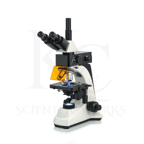
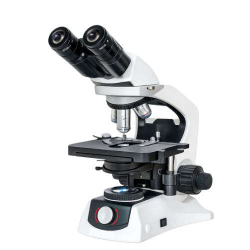
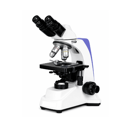
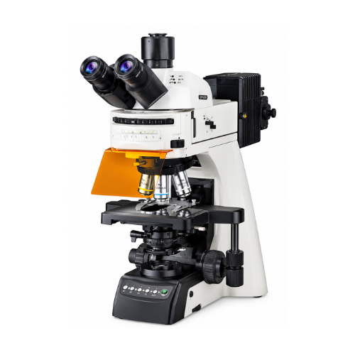
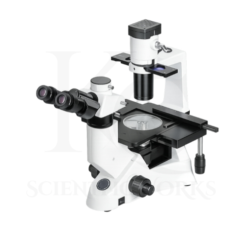
The Fluorescent Microscope KCX-160 is designed for microscopic examination of biological specimens using fluorescence and conventional transmitted-light observation techniques.
In fluorescence microscopy, a specimen is prepared using suitable fluorescent dyes, labels or naturally fluorescent materials.
The microscope illuminates the specimen with excitation light selected for the fluorescent material.
The fluorescent material absorbs the excitation light and emits light at a different wavelength.
An appropriate filter arrangement separates the emitted fluorescence from unwanted excitation light, allowing selected structures to be observed.
This technique can help researchers examine labelled cells, tissues, microorganisms and biological structures.
The KCX-160 also supports conventional bright-field observation for suitable prepared specimens.
Its optional phase-contrast configuration may be used for observing appropriate transparent specimens when the required phase-contrast optical components are installed.
The microscope's mechanical stage, coaxial focusing system and adjustable illumination controls allow users to position and examine specimens during laboratory investigations.
With compatible digital imaging accessories, the trinocular configuration can support image capture and research documentation.
Applications
1. Fluorescence Microscopy
The KCX-160 Fluorescent Microscope is suitable for examining compatible fluorescent specimens using the installed excitation and emission filter arrangement.
2. Cell Biology Research
Cell biology laboratories can use the microscope to examine appropriately prepared and fluorescently labelled cellular structures.
3. Biotechnology Laboratories
The microscope supports suitable fluorescence-based observation and biological specimen examination in biotechnology research environments.
4. Microbiology Laboratories
Microbiology laboratories can use the KCX-160 for observing appropriately prepared microorganisms using suitable bright-field or fluorescence methods.
5. Immunofluorescence Studies
The microscope can support immunofluorescence-based observation when the installed filter set is compatible with the fluorescent label being used.
6. Histology Laboratories
The KCX-160 can be used to examine suitable prepared tissue sections using bright-field or compatible fluorescence techniques.
7. Molecular Biology Research
The microscope can support observation of fluorescently labelled biological specimens in molecular biology research.
8. Pharmaceutical Research
Pharmaceutical laboratories can use the microscope for appropriate cellular and fluorescence-based research applications.
9. Medical Research Laboratories
The microscope is suitable for research-oriented examination of prepared tissues, cells and compatible fluorescent specimens.
10. University Research Departments
Universities can use the KCX-160 for advanced biological microscopy training and scientific research.
11. Scientific Research Institutes
Research institutions can use the microscope for suitable fluorescence observation and laboratory specimen analysis.
12. Biological Education
The microscope supports teaching of fluorescence microscopy, optical magnification and advanced biological specimen examination.
13. Tissue Examination
The KCX-160 can be used to observe appropriately prepared tissue specimens using compatible optical observation modes.
14. Cellular Imaging
With suitable fluorescent labels and filter arrangements, the microscope can support visualization of selected cellular components.
15. Laboratory Training
The microscope is useful for teaching illumination adjustment, objective selection, focusing, fluorescence filter use and specimen positioning.
16. Digital Research Documentation
The trinocular configuration can support compatible digital imaging equipment for recording selected microscope images.
Working Principle
The Fluorescent Microscope KCX-160 operates using the principles of optical magnification, fluorescence excitation and emission, and transmitted-light microscopy.
In fluorescence mode, a prepared specimen containing suitable fluorescent material is positioned on the mechanical stage.
The LED fluorescence illumination system produces excitation light.
The excitation light passes through the appropriate illumination optics and filter arrangement before reaching the specimen.
Fluorescent molecules within the specimen absorb light at suitable excitation wavelengths.
After excitation, the fluorescent material emits light at a longer wavelength.
The microscope objective collects the emitted fluorescence and forms a magnified optical image.
The fluorescence filter arrangement helps separate the desired emitted light from the excitation illumination.
The resulting image is observed through the binocular or trinocular optical system.
The coaxial focusing mechanism allows users to adjust image sharpness, while the mechanical stage supports controlled specimen positioning.
For bright-field observation, transmitted light from the 10W LED illumination system passes through the specimen and is collected by the selected objective lens.
The microscope therefore supports different observation methods according to the installed optical and illumination configuration.
Fluorescence Observation Process
Specimen Preparation: A suitable specimen is prepared with a compatible fluorescent label or naturally fluorescent material.
Excitation Illumination: The LED fluorescence light source produces excitation light.
Optical Filtering: The excitation filter arrangement selects suitable illumination wavelengths.
Fluorescence Emission: Fluorescent material emits light after excitation.
Emission Filtering: The filter arrangement separates desired fluorescence from unwanted excitation light.
Optical Magnification: The objective lens forms a magnified image.
Observation: The image is viewed through the eyepieces or a compatible camera system.
Magnification Combinations
| Objective Lens | Numerical Aperture | Eyepiece | Total Optical Magnification |
|---|
| Plan Achromatic 4X | 0.10 | WF10X | 40X |
| Plan Achromatic 10X | 0.25 | WF10X | 100X |
| Plan Achromatic 40X | 0.65 | WF10X | 400X |
| Plan Achromatic 100X | 1.30 | WF10X | 1000X |
Optional Magnification Configurations
| Optional Magnification | Availability |
|---|
| 600X | Optional Optical Configuration |
| 800X | Optional Optical Configuration |
The exact objective and eyepiece combinations for optional magnifications should be confirmed from the final product configuration.
Fluorescence Imaging System
| Parameter | Specification |
|---|
| Fluorescence Illumination | LED-Based |
| Fluorescence Intensity | Adjustable |
| Fluorescence Filter | Single Immunofluorescence Filter |
| Specified LED Life | Up to 60,000 Hours |
| Observation Method | Fluorescence Microscopy |
| Compatible Fluorophores | Depends on Installed Filter Set |
| Camera Integration | Supported with Compatible Trinocular Configuration |
| Additional Filter Channels | Not Specified |
| Fluorescence Camera Resolution | Not Specified |
| Digital Recording | Depends on Installed Camera and Software |
Microscope Compatibility and Usage
The KCX-160 Fluorescent Microscope is intended for laboratory observation of suitable prepared biological and fluorescent specimens.
It can be used in biotechnology laboratories, research institutes, universities, pharmaceutical research centers and scientific training institutions.
For routine bright-field observation, the specimen should be placed on the mechanical stage and illuminated using the transmitted LED light source.
The operator can select the required objective lens and adjust coarse and fine focusing until a clear image is obtained.
For fluorescence observation, the specimen must be compatible with the installed excitation light source and fluorescence filter arrangement.
The fluorescence illumination intensity should be adjusted according to the specimen and observation requirements.
The microscope should be used with appropriate fluorescence laboratory practices to minimize unnecessary exposure of light-sensitive specimens.
The trinocular configuration may be connected to compatible digital imaging equipment for specimen documentation.
For phase-contrast observation, suitable phase objectives and condenser components must be installed.
The microscope should be operated according to its final supplied technical configuration and laboratory safety instructions.