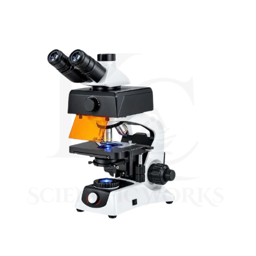
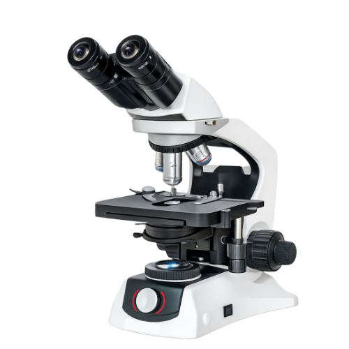
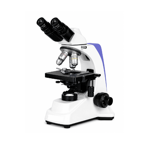
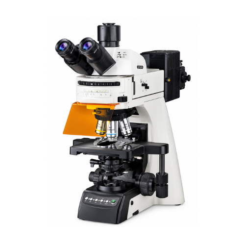
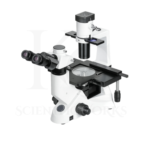
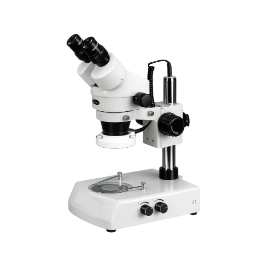
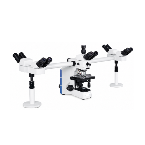
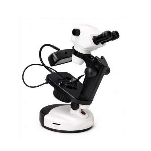
The Fluorescent Microscope KCX-162 is designed to support advanced microscopic examination of biological specimens using both fluorescence and conventional bright-field observation techniques.
Fluorescence microscopy is used when selected biological structures are labelled with fluorescent materials or exhibit suitable natural fluorescence.
The microscope illuminates the specimen with excitation light from its 100W HBO mercury lamp through an appropriate optical filter arrangement.
Fluorescent molecules within the specimen absorb excitation light and emit light at characteristic wavelengths.
The microscope collects the emitted light and directs it through the optical system for magnified observation.
The multi-channel fluorescence system allows users to select among blue, green and UV excitation configurations according to the fluorescent labels and filter sets available.
The microscope's plan-achromatic objectives support examination of suitable biological structures, while its trinocular head enables integration with compatible digital imaging equipment.
Conventional bright-field microscopy can also be used for examining prepared biological specimens without fluorescence illumination.
The KCX-162 is therefore intended for immunofluorescence research, cell biology, microbiology, pathology research, biotechnology and scientific education.
Applications
1. Immunofluorescence Research
The KCX-162 Fluorescent Microscope can be used to examine biological specimens labelled with compatible fluorescent antibodies.
Its multi-channel fluorescence configuration supports appropriate immunofluorescence observation workflows.
2. Cell Biology Research
Cell biology laboratories can use the microscope to examine suitable cellular structures, morphology and fluorescent markers.
3. Pathology Research
The microscope can support research-oriented examination of appropriately prepared tissue and cellular specimens.
4. Microbiology Laboratories
Microbiology researchers can use the KCX-162 for suitable fluorescence-based observation of labelled microorganisms.
5. Fluorescent Staining Studies
The microscope supports examination of specimens prepared using compatible fluorescent staining methods.
6. Cellular Morphology
The KCX-162 can be used to examine suitable cellular structures through bright-field and fluorescence microscopy.
7. Molecular Biology Research
The microscope can support observation of fluorescently labelled biological materials used in suitable molecular biology investigations.
8. Pharmaceutical Research
Pharmaceutical research laboratories can use the microscope for appropriate cellular and fluorescence-based investigations.
9. Biotechnology Laboratories
The microscope is suitable for research involving fluorescent markers, prepared biological specimens and compatible microscopy assays.
10. Medical Research Laboratories
The KCX-162 can support research-oriented observation of biological samples in medical research environments.
11. University Research Departments
Universities can use the microscope for advanced biological microscopy teaching and laboratory research.
12. Scientific Research Institutes
The microscope supports suitable fluorescence-based investigations in research institutions.
13. Biological Specimen Examination
The KCX-162 can be used to examine appropriately prepared tissues, cells and microorganisms.
14. Diagnostic Method Development
Research laboratories can use the microscope to investigate fluorescence-based laboratory methods.
Use for actual clinical diagnosis requires appropriate assay validation, regulatory compliance and approved operating procedures.
15. Fluorescence-Based Microbial Studies
The microscope may support laboratory research involving fluorescently labelled microbial specimens and compatible detection methods.
16. Digital Microscopy Documentation
The trinocular head can be integrated with compatible cameras for recording selected microscope images.
17. Advanced Scientific Education
The microscope is suitable for teaching fluorescence principles, optical imaging and laboratory microscopy techniques.
18. Specialized Laboratory Research
The KCX-162 supports appropriate research applications requiring multiple fluorescence excitation ranges and high-magnification observation.
Working Principle
The Fluorescent Microscope KCX-162 operates using the principles of optical magnification, fluorescence excitation, fluorescence emission and wavelength-selective optical filtering.
A suitably prepared biological specimen is placed on the microscope's mechanical stage.
The 100W HBO mercury lamp produces light containing multiple spectral components.
An excitation filter selects the wavelength range required to illuminate the fluorescent material within the specimen.
The selected excitation light is directed toward the specimen through the fluorescence illumination optics.
Fluorescent molecules absorb the excitation light and enter an excited electronic state.
As these molecules return to a lower energy state, they emit light, typically at a longer wavelength than the excitation light.
The microscope objective collects the emitted fluorescence.
The fluorescence filter arrangement separates the desired emitted light from unwanted excitation illumination.
The resulting optical image passes through the microscope's infinity-corrected optical system and is observed through the trinocular viewing head.
A compatible camera can also be used for digital image acquisition when suitable accessories are installed.
Fluorescence Observation Process
Specimen Preparation: A compatible biological specimen is prepared using suitable fluorescent labels or naturally fluorescent material.
Excitation Light Generation: The 100W HBO mercury lamp produces illumination.
Excitation Filtering: The selected fluorescence filter isolates the required excitation wavelength range.
Specimen Excitation: Fluorescent molecules absorb the excitation light.
Fluorescence Emission: The fluorescent material emits light at characteristic wavelengths.
Emission Filtering: The optical filter arrangement separates desired fluorescence from unwanted excitation light.
Optical Magnification: The objective and microscope optical system form a magnified image.
Observation and Documentation: The image is viewed through the eyepieces or captured using a compatible camera.
Magnification System
| Parameter | Specification |
|---|
| Optical System | Infinity-Corrected |
| Eyepiece | WF10X |
| Objective Range | 4X–100X |
| Standard Magnification Range | 40X–1000X |
| Specified Maximum Magnification | Up to 2500X |
| High-Power Objective | 100X Oil Immersion |
| 100X Objective Numerical Aperture | 1.25 |
Standard Optical Magnification Examples
| Objective | Eyepiece | Total Magnification |
|---|
| 4X | 10X | 40X |
| 10X* | 10X | 100X |
| 40X* | 10X | 400X |
| 100X Oil Immersion | 10X | 1000X |
10X and 40X objectives are illustrative intermediate magnifications within the stated objective range; their exact specifications should be confirmed for the KCX-162.
The 2500X specification requires a compatible additional optical configuration. Higher nominal magnification does not necessarily provide greater optical resolution.
Fluorescence Filter System
| Fluorescence Channel | Specified Wavelength Range |
|---|
| Blue | 420–485 nm |
| Green | 480–550 nm |
| UV | 330–400 nm |
These wavelength ranges are taken from the supplied reference. They do not, by themselves, establish complete fluorescence filter-set compatibility.
The excitation filter, dichroic mirror, emission filter and fluorescent label must be matched for the intended application.
Digital Imaging Compatibility
| Parameter | Specification |
|---|
| Viewing Head | Trinocular |
| Camera Attachment | Supported with Compatible Adapter |
| Digital Image Capture | Configuration-Dependent |
| Camera Resolution | Not Specified |
| Image Measurement Software | Not Specified |
| Live Computer Display | Depends on Installed Camera |
| Video Recording | Depends on Installed Imaging System |
Microscope Compatibility and Usage
The KCX-162 Fluorescent Microscope is intended for observation of suitable prepared biological specimens in scientific and research laboratories.
It can be used in universities, biotechnology laboratories, microbiology departments, pharmaceutical research centers and specialized scientific institutions.
For bright-field observation, the specimen is placed on the mechanical stage and the transmitted-light illumination system is selected.
For fluorescence observation, the operator must select a compatible filter configuration and use a specimen prepared for the relevant excitation and emission wavelengths.
The selected objective lens and illumination settings should be appropriate for the specimen.
The microscope's trinocular head may be integrated with compatible digital imaging accessories for documentation and research analysis.
The 100W HBO high-pressure mercury lamp must be operated only with its specified power supply, enclosed lamp housing, protective optical components and manufacturer-approved safety procedures. Users must not look directly at the lamp or unfiltered UV illumination. Lamp handling, replacement and disposal require appropriate trained personnel and procedures.
The microscope should be used according to its final technical configuration, laboratory safety instructions and applicable regulatory requirements.