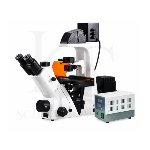
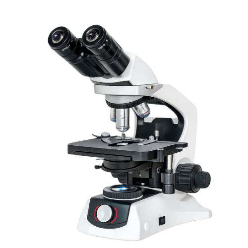
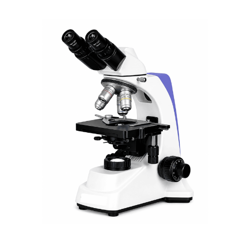
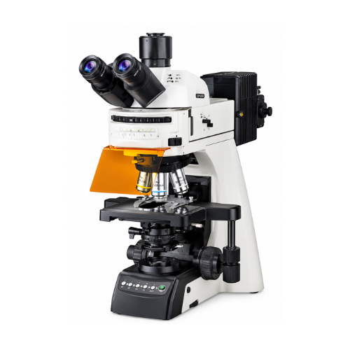
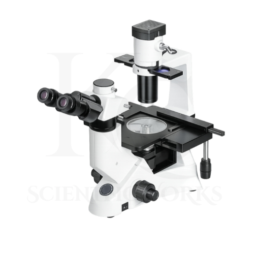
The Inverted Fluorescence Microscope KCX-166 is designed for high-resolution observation of suitable biological specimens using inverted optical microscopy and multiple illumination techniques.
Its inverted design allows specimens to be viewed from beneath the stage, making it particularly useful for examining compatible cell cultures and biological samples in laboratory vessels.
The microscope supports conventional bright-field observation, phase-contrast microscopy, fluorescence imaging and DIC configurations.
Bright-field microscopy provides general transmitted-light observation of suitable specimens.
Phase-contrast microscopy can improve visibility of transparent and unstained specimens when appropriate phase objectives and condenser components are installed.
Fluorescence microscopy allows suitably labelled biological structures to be observed using selected excitation and emission filter arrangements.
DIC microscopy can provide contrast in suitable transparent specimens when the required DIC optical components are installed.
The microscope's digital imaging system allows images to be captured and processed using compatible software.
Time-lapse imaging can support observation of changes in suitable specimens over time.
Z-stack acquisition can support imaging at different focal positions when the required hardware and software functions are available.
The KCX-166 is therefore intended for advanced biological research, fluorescence observation, cell culture studies and scientific image documentation.
Applications
1. Cell Biology Research
The KCX-166 Inverted Fluorescence Microscope is suitable for observing compatible cell cultures and fluorescently labelled cellular structures.
2. Live-Cell Imaging
The inverted optical arrangement supports observation of living cells in suitable culture vessels.
Long-duration experiments may require temperature, humidity and gas-control accessories.
3. Molecular Biology Research
The microscope can support observation of fluorescently labelled biological materials used in molecular biology investigations.
4. Cancer Research
Research laboratories can use the microscope for suitable cell-based studies and fluorescence imaging applications related to cancer biology.
5. Immunofluorescence Studies
The DAPI, FITC, TRITC and CY5 filter configurations support observation of compatible fluorescently labelled specimens.
6. Microbiology Laboratories
The microscope can be used for observing suitable microorganisms and prepared biological specimens using compatible microscopy techniques.
7. Stem Cell Research
The KCX-166 can support observation of suitable stem cell cultures and fluorescently labelled cellular structures.
8. Pharmaceutical Research
Pharmaceutical laboratories can use the microscope for appropriate cell-based imaging and fluorescence research applications.
9. Drug Discovery Research
The microscope can support microscopy-based observation in suitable experimental drug discovery workflows.
10. Biotechnology Laboratories
The KCX-166 is suitable for fluorescence imaging and observation of compatible biological specimens in biotechnology research.
11. Histopathology Research
The microscope can support research-oriented examination of suitable prepared tissue specimens, subject to compatibility with the inverted stage and optical arrangement.
12. Biomedical Research
Biomedical research institutions can use the microscope for appropriate fluorescence and cell imaging investigations.
13. IVF Research Laboratories
The inverted microscope configuration may support suitable reproductive biology research involving compatible specimens and laboratory vessels.
Clinical embryology use requires equipment and procedures validated for that purpose.
14. Medical Universities
Medical universities can use the microscope for advanced biological microscopy education and research.
15. Scientific Research Institutes
Research institutes can use the KCX-166 for fluorescence observation, digital imaging and suitable biological experiments.
16. Clinical Laboratory Research
The microscope may be used for research and method-development activities involving prepared biological specimens.
Clinical diagnostic use requires appropriate validation and regulatory compliance.
17. Fluorescent Staining Studies
The microscope supports observation of compatible fluorescently stained specimens using the installed filter sets.
18. Time-Lapse Microscopy
The digital imaging system supports time-lapse acquisition for suitable experiments.
19. Z-Stack Imaging Research
The microscope can support Z-stack acquisition when the required focusing hardware and software components are installed.
20. Digital Image Analysis
The supplied software configuration supports image measurement and analysis of compatible microscope images.
Working Principle
The Inverted Fluorescence Microscope KCX-166 operates using the principles of inverted optical microscopy, optical magnification, fluorescence excitation and emission, and digital image acquisition.
Unlike upright microscopes, an inverted microscope positions its objective lenses below the specimen stage.
A compatible biological specimen is placed in a suitable culture vessel or laboratory container on the stage.
The microscope objective collects light from the specimen through the bottom of the vessel.
In bright-field mode, transmitted illumination passes through the specimen and is collected by the objective.
In phase-contrast mode, compatible phase optical components convert selected phase differences in transmitted light into visible intensity differences.
In fluorescence mode, excitation light from the fluorescence illumination system is directed toward the specimen through the appropriate filter arrangement.
Fluorescent labels absorb excitation light and emit light at characteristic wavelengths.
The microscope's emission filters help separate the desired fluorescent signal from unwanted excitation illumination.
The resulting image is magnified by the infinity-corrected optical system and observed through the eyepieces or acquired through the digital camera port.
In DIC mode, suitable polarization and interference optics create contrast from optical path differences in compatible specimens.
The digital CMOS camera converts the optical image into electronic image data.
The imaging software can then support live observation, image capture, measurement and compatible advanced imaging functions.
Fluorescence Imaging Process
Specimen Preparation: A compatible biological specimen is prepared using suitable fluorescent labels.
Specimen Placement: The specimen is positioned in a compatible vessel on the mechanical stage.
Excitation Illumination: The fluorescence system produces light for the selected fluorescence channel.
Optical Filtering: The excitation filter selects suitable wavelengths.
Fluorescence Emission: Fluorescent molecules emit light after excitation.
Image Formation: The objective and infinity-corrected optical system form a magnified image.
Digital Acquisition: The scientific CMOS camera captures the optical image.
Image Processing: The imaging software supports compatible documentation and analysis functions.
Magnification System
| Objective | Eyepiece | Nominal Optical Magnification |
|---|
| 10X Long Working Distance | WF10X | 100X |
| 20X Long Working Distance | WF10X | 200X |
| 40X Long Working Distance | WF10X | 400X |
| 60X Long Working Distance | WF10X | 600X |
The listed magnifications are calculated from the supplied objective and eyepiece values. Actual optical performance depends on objective specifications, numerical aperture and the final microscope configuration.
Fluorescence Filter System
| Filter Set | Intended Fluorescence Channel |
|---|
| DAPI | DAPI-Compatible Fluorescence |
| FITC | FITC-Compatible Fluorescence |
| TRITC | TRITC-Compatible Fluorescence |
| CY5 | CY5-Compatible Fluorescence |
The exact excitation and emission wavelengths must be confirmed from the installed filter-set specifications.
Digital Imaging System
| Parameter | Specification |
|---|
| Camera Type | Scientific CMOS |
| Specified Camera Resolution | 25 MP |
| Specified Image Resolution | 4800 × 3600 Pixels |
| Maximum Frame Rate | Up to 60 fps |
| USB Interface | USB 3.2 Gen 2 |
| Display Interface | HDMI |
| Wireless Connectivity | Wi-Fi |
| Imaging Software | Scientific Imaging and Measurement |
| Time-Lapse Imaging | Supported |
| Z-Stack Imaging | Configuration-Dependent |
| Image Stitching | Software-Dependent |
| AI-Assisted Analysis | Software-Dependent |
| Computer | Intel Core i7 |
| RAM | 8 GB |
| Storage | 1 TB SSD |
| Operating System | Windows 10 / Windows 11 |
Microscope Compatibility and Usage
The KCX-166 Inverted Fluorescence Microscope is designed for observation of suitable biological specimens, cell cultures and fluorescently labelled samples.
It can be used in biotechnology laboratories, biomedical research centers, universities, pharmaceutical research facilities and scientific institutions.
For routine observation, the specimen should be placed in a compatible laboratory vessel on the mechanical stage.
The appropriate objective lens should be selected according to the specimen and observation requirements.
The white LED illumination system can be used for compatible transmitted-light observation.
For fluorescence imaging, the selected fluorescent label must be compatible with the installed excitation and emission filter arrangement.
Phase-contrast observation requires suitable phase objectives and condenser components.
DIC observation requires the appropriate DIC optical accessories.
The integrated camera system can be used for image acquisition and documentation through compatible software.
For extended live-cell imaging, suitable environmental control equipment may be required to maintain specimen conditions.
The microscope should be operated according to its final supplied technical configuration and laboratory safety instructions.