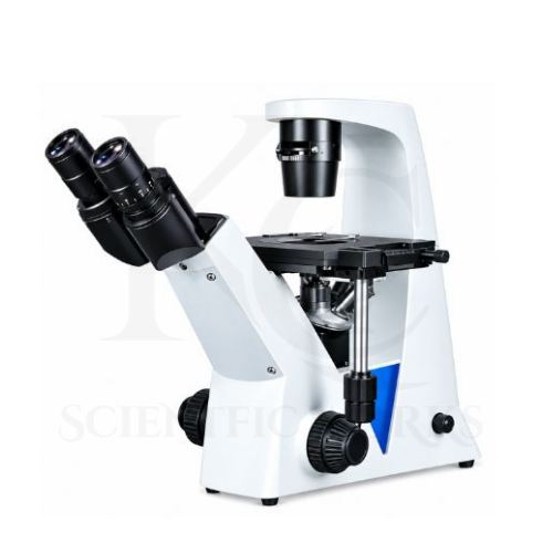
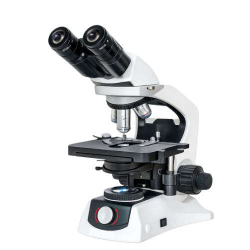
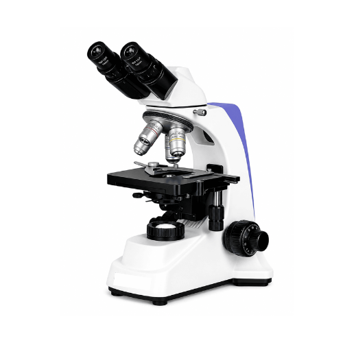
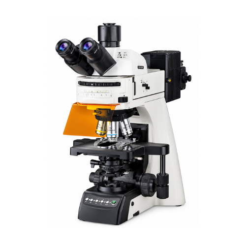
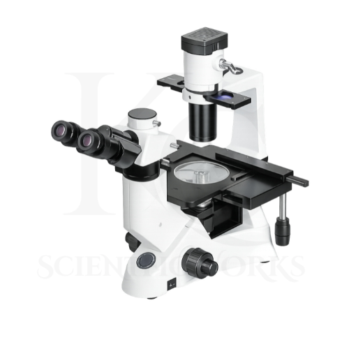
The Inverted Phase Contrast Microscope KCX-171 is designed primarily for tissue culture, cell culture and live-cell observation.
Its inverted optical configuration positions the objective lenses below the specimen stage.
This allows researchers to observe suitable biological specimens through the transparent bottom of compatible culture dishes, flasks and other laboratory vessels.
The microscope supports both bright-field and phase-contrast observation.
Bright-field microscopy is useful for general observation of suitably prepared specimens.
Phase-contrast microscopy improves the visibility of transparent and low-contrast specimens by converting optical phase differences into visible intensity variations.
This makes phase contrast useful for examining suitable living cells without routine staining.
The microscope's long working distance objectives provide clearance for compatible culture vessels.
The mechanical stage and attachable holders support specimen positioning.
The trinocular observation head allows a compatible camera to be attached for image capture and documentation.
The KCX-171 is therefore intended for cell culture monitoring, tissue culture research, cell morphology examination, biotechnology studies and advanced biological microscopy education.
Applications
1. Tissue Culture Laboratories
The KCX-171 Inverted Phase Contrast Microscope is suitable for routine observation of compatible tissue culture specimens.
2. Cell Culture Laboratories
The microscope supports observation of living cells in suitable culture dishes and flasks.
3. Cell Biology Research
Researchers can use the microscope to examine cellular morphology and other visible characteristics.
4. Live-Cell Observation
The inverted phase-contrast configuration is useful for observing suitable living cells without routine staining.
5. Biotechnology Research
The microscope supports microscopy-based observation in biotechnology laboratories.
6. Molecular Biology Research
The KCX-171 can support examination of suitable biological specimens used in molecular biology studies.
7. Microbiology Laboratories
The microscope may be used for observation of compatible microorganisms and cell cultures.
8. Pharmaceutical Research
Pharmaceutical laboratories can use the microscope for suitable cell-based research and observation workflows.
9. Drug Discovery Research
The microscope can support observation of cell cultures used in appropriate drug discovery studies.
10. Cancer Research
Research laboratories can use the KCX-171 for suitable cell culture observation associated with cancer biology investigations.
11. Stem Cell Research
The microscope supports observation of compatible stem cell cultures and cellular morphology.
12. IVF and Reproductive Biology Research
The inverted microscope configuration may support suitable reproductive biology research involving compatible specimens.
Clinical embryology use requires appropriate validation and specialized operating procedures.
13. Immunology Research
The microscope can support observation of suitable cell cultures used in immunology research.
14. Virology Research
The KCX-171 can support observation of compatible cell cultures used in virology research workflows.
15. Biomedical Research
Biomedical laboratories can use the microscope for appropriate cell-based observation and documentation.
16. Life Science Laboratories
The microscope is suitable for biological microscopy in life science research and teaching laboratories.
17. Medical Research Institutions
Medical research institutions can use the KCX-171 for research-oriented observation of suitable cell cultures.
18. Universities and Colleges
Universities can use the microscope for tissue culture education, cell biology practicals and laboratory research.
19. Research Institutes
Scientific institutions can use the microscope for advanced cell observation and phase-contrast microscopy.
20. Cell Culture Monitoring
The microscope can support visual monitoring of cell morphology, density and visible culture characteristics.
Typical Observation and Research Areas
| Observation Area | Typical Purpose |
|---|
| Living Cell Cultures | Routine visual observation of suitable living cells |
| Tissue Culture Samples | Examination of cultured biological specimens |
| Mammalian Cells | Observation of morphology and culture characteristics |
| Cell Morphology | Examination of cell shape and visible structures |
| Cell Growth | Monitoring visible changes in cell cultures |
| Cell Attachment | Observing attachment characteristics of suitable adherent cells |
| Cell Proliferation | Visual monitoring of changes in cell populations |
| Cell Density | Qualitative assessment of culture coverage |
| Cell Viability | Preliminary visual observations; quantitative viability requires validated assays |
| Microorganisms | Observation of compatible microbial specimens |
| Culture Contamination | Visual screening for certain contamination signs; not a substitute for validated testing |
| Drug-Response Studies | Observation of visible cellular changes during appropriate research |
| Cellular Morphology Changes | Comparison of visible specimen characteristics |
| Culture Vessel Observation | Viewing suitable specimens through compatible vessel bottoms |
Working Principle
The Inverted Phase Contrast Microscope KCX-171 operates using the principles of inverted optical microscopy, transmitted-light illumination and phase-contrast image formation.
Unlike conventional upright microscopes, an inverted microscope positions its objective lenses beneath the specimen stage.
A compatible culture vessel containing the specimen is placed on the microscope stage.
Light from the 6V / 30W halogen illumination system passes through the condenser and specimen.
The objective lens beneath the specimen collects the transmitted light.
In bright-field observation, differences in specimen absorption and scattering produce visible image contrast.
In phase-contrast observation, the condenser annulus and compatible phase objective work together to convert optical phase differences into visible intensity differences.
This improves the visibility of many transparent and low-contrast specimens.
The microscope's infinity plan optical system forms a magnified image for observation through the eyepieces.
The trinocular head can also direct light toward a compatible camera for digital imaging.
The coaxial coarse and fine focusing controls allow the user to adjust image sharpness.
Phase Contrast Observation Process
Specimen Preparation: A suitable biological specimen is prepared in a compatible culture vessel.
Specimen Placement: The vessel is positioned on the working stage using an appropriate holder.
Illumination: The transmitted halogen light source provides illumination through the condenser.
Objective Selection: The required long working distance objective is selected.
Phase Annulus Selection: The matching phase-contrast annulus is selected for phase observation.
Focusing: Coarse and fine focusing controls are adjusted to obtain a clear image.
Image Observation: The specimen is examined through the eyepieces.
Digital Documentation: A compatible camera may be used for image capture when installed.
Magnification System
| Objective | Eyepiece | Total Optical Magnification |
|---|
| 10X Infinity Plan LWD Semi-APO | WF10X | 100X |
| 20X Infinity Plan LWD Semi-APO | WF10X | 200X |
| 40X Infinity Plan LWD Semi-APO | WF10X | 400X |
| 20X Infinity Phase Contrast LWD Semi-APO | WF10X | 200X |
The microscope's standard nominal optical magnification range is 100X–400X.
Phase Contrast Optical System
| Parameter | Specification |
|---|
| Observation Mode | Phase Contrast |
| Optical System | Infinity Plan |
| Dedicated Phase Objective | LWD Semi-APO FL 20X |
| Phase Annular Spots | 10X and 20X/40X Configurations |
| Condenser | LWD / ELWD |
| Condenser N.A. | 0.3 |
| Phase Contrast Slide Slot | Included |
| Main Purpose | Observation of Suitable Transparent and Low-Contrast Specimens |
Stage and Culture Vessel Compatibility
| Parameter | Specification |
|---|
| Stage Type | Fixed with Attachable Components |
| Stage Dimensions | 210 × 241 mm |
| Specified Working Area | ≥140 × 180 mm |
| Round Slide Accommodation | Ø110 mm |
| Culture Dish Holder | 54 mm |
| Condenser Working Distance | 72 mm with Condenser |
| Clearance Without Condenser | 195 mm |
| Intended Specimens | Compatible Culture Dishes, Flasks and Laboratory Vessels |
The compatibility of individual culture vessels depends on their dimensions, bottom thickness, optical properties and the installed holder.
Digital Imaging Compatibility
| Parameter | Specification |
|---|
| Viewing Head | Trinocular |
| Light Path | E100 / E20 |
| Camera Port | Supported |
| Digital Camera | Compatible Camera Can Be Attached |
| Camera Resolution | Not Specified |
| Image Capture | Depends on Installed Camera |
| Video Recording | Depends on Installed Camera |
| Imaging Software | Not Specified |
| Computer Connection | Depends on Selected Camera System |
Microscope Compatibility and Usage
The KCX-171 Inverted Phase Contrast Microscope is designed for observation of suitable cell cultures, tissue culture samples and other biological specimens.
It can be used in cell culture laboratories, biotechnology research centers, universities, pharmaceutical research facilities and scientific institutions.
For routine observation, the specimen should be placed in a compatible culture vessel on the working stage.
The appropriate long working distance objective should be selected according to the specimen and vessel configuration.
Bright-field observation can be used for general specimen examination.
For phase-contrast observation, the appropriate phase objective and matching condenser annulus must be selected and aligned.
The microscope's focusing controls allow adjustment of image sharpness.
The trinocular port can be used for compatible digital camera integration.
For extended live-cell observation, additional environmental control accessories may be necessary to maintain suitable specimen conditions.
The microscope should be operated according to its final supplied technical configuration and laboratory safety instructions.