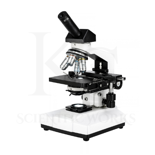
The Monocular Microscope KCX-135 is developed for observing microscopic structures in suitable biological and laboratory specimens.
It allows students, educators, laboratory technicians and researchers to examine specimens that cannot be adequately distinguished with the unaided eye.
The microscope incorporates plan achromatic objectives with magnifications of 4X, 10X, 40X and 100X.
Combined with the WF10X eyepiece, these objectives provide nominal magnifications of 40X, 100X, 400X and 1000X.
The LED illumination system provides transmitted light for observing suitable prepared specimens.
The Abbe condenser and adjustable iris diaphragm help control illumination.
The mechanical stage supports controlled specimen positioning.
Separate coarse and fine focusing controls allow users to obtain a clear image.
The microscope is suitable for biology education, laboratory training, microbiology studies, prepared tissue observation and routine scientific examination.
Applications
1. School Science Laboratories
The KCX-135 Monocular Microscope supports biology practical classes and introductory laboratory microscopy.
2. Colleges and Universities
The microscope is suitable for educational demonstrations and practical laboratory training.
3. Biology Laboratories
The KCX-135 can be used to examine suitable prepared plant and animal specimens.
4. Microbiology Laboratories
The microscope supports observation of appropriately prepared microorganisms.
5. Biotechnology Laboratories
The KCX-135 can support routine microscopy activities in biotechnology education and research.
6. Research Institutes
Research laboratories can use the microscope for examination of compatible biological specimens.
7. Medical Training Centers
The microscope supports educational observation of prepared biological and pathology slides.
8. Pathology Teaching Laboratories
The KCX-135 is suitable for observing prepared tissue slides in pathology education.
9. Clinical Laboratory Training
The microscope can be used for microscopy instruction involving suitable prepared clinical teaching specimens.
10. Plant Cell Observation
Students can examine visible structures in appropriately prepared plant cell specimens.
11. Animal Cell Observation
The microscope supports examination of suitable prepared animal cell slides.
12. Tissue Examination
The KCX-135 can be used for observation of prepared biological tissue sections.
13. Prepared Blood Smear Observation
The microscope supports educational observation of appropriately prepared blood smears.
14. Bacterial Observation
The 100X oil immersion objective supports examination of appropriately stained bacterial specimens.
15. Educational Demonstrations
Teachers can demonstrate microscope components, magnification and focusing techniques.
16. Laboratory Skill Development
Students can practice specimen handling, focusing and microscope operation.
17. Scientific Experiments
The microscope supports suitable educational experiments involving optical microscopy.
18. Routine Laboratory Observation
The KCX-135 is suitable for general observation of compatible prepared specimens.
19. Science Training Programs
The microscope can be used in educational and laboratory training institutions.
20. Biological Research
The microscope supports appropriate bright-field microscopy applications in biological research.
Typical Observation Areas
| Observation Area | Typical Purpose |
|---|
| Plant Cells | Observation of visible cellular structures |
| Animal Cells | Examination of prepared cell specimens |
| Tissue Sections | Observation of prepared biological tissues |
| Blood Smears | Educational examination of stained blood cells |
| Bacterial Slides | Observation of appropriately stained bacteria |
| Fungal Specimens | Examination of suitable prepared fungal structures |
| Microorganisms | Observation of compatible microscopic organisms |
| Leaf Sections | Examination of plant tissue organization |
| Stem Sections | Observation of plant tissue structures |
| Prepared Pathology Slides | Teaching and research observation |
| Educational Slides | Microscope handling and focusing practice |
| Biological Specimens | Routine bright-field microscopy |
Working Principle
The Monocular Microscope KCX-135 operates using the principle of compound optical magnification.
A suitable prepared specimen is positioned on the mechanical stage.
The LED illumination system provides light that passes through the specimen.
The Abbe condenser concentrates and directs light toward the specimen.
The selected objective lens collects transmitted light and forms a magnified intermediate image.
The WF10X eyepiece further magnifies the image for the observer.
The total nominal magnification is calculated by multiplying the objective magnification by the eyepiece magnification.
The coarse and fine focusing controls are used to obtain a clear image.
For suitable high-magnification observations, the 100X oil immersion objective is used with compatible immersion oil according to laboratory procedures.
Basic Microscope Observation Process
Microscope Placement: Position the KCX-135 on a stable laboratory bench.
Specimen Preparation: Prepare or select a suitable microscope slide.
Slide Positioning: Place the slide on the mechanical stage.
Illumination: Switch on the LED light source and adjust brightness.
Objective Selection: Begin with the 4X objective.
Coarse Focusing: Carefully adjust the coarse focus to locate the specimen.
Fine Focusing: Adjust the fine focus for image clarity.
Magnification Change: Select a higher-power objective when needed.
Condenser Adjustment: Adjust the condenser and iris diaphragm as appropriate.
Oil Immersion Observation: Use the 100X oil immersion objective with suitable immersion oil when required.
Specimen Examination: Observe and document visible specimen characteristics.
Magnification System
| Objective | Eyepiece | Total Optical Magnification |
|---|
| 4X Plan Achromatic | WF10X | 40X |
| 10X Plan Achromatic | WF10X | 100X |
| 40X Plan Achromatic (SL) | WF10X | 400X |
| 100X Plan Achromatic Oil | WF10X | 1000X |
Standard nominal magnification range: 40X–1000X.
Optical System
| Parameter | Specification |
|---|
| Microscope Type | Monocular Compound |
| Eyepiece | WF10X / FN18 |
| Objectives | 4X, 10X, 40X (SL), 100X Oil |
| Objective Correction | Plan Achromatic |
| Nosepiece | Quadruple Revolving |
| Condenser | Abbe |
| Condenser N.A. | 1.25 |
| Diaphragm | Iris |
| Illumination | LED |
| Brightness Control | Adjustable |
Mechanical Stage and Focusing System
| Parameter | Specification |
|---|
| Stage Type | Mechanical |
| Stage Size | 120 × 125 mm |
| Focusing | Separate Coarse and Fine |
| Head Inclination | 45° |
| Head Rotation | 360° |
| Body Construction | Heavy-Duty Metal |
Educational and Laboratory Benefits
The KCX-135 Monocular Microscope supports practical scientific learning and routine laboratory observation.
It helps users understand:
Compound optical magnification
Objective and eyepiece functions
Microscope focusing techniques
Mechanical stage operation
Condenser and iris diaphragm adjustment
Bright-field microscopy
Biological specimen preparation
Oil immersion microscopy
Plant and animal cell observation
Microorganism examination
Routine laboratory instrument handling
Scientific observation and documentation
Microscope Compatibility and Usage
The KCX-135 Monocular Microscope is intended for observation of suitable prepared microscope slides.
It can be used in educational institutions, biology laboratories, microbiology training centers and research facilities.
The microscope should be positioned on a stable surface.
Specimens should be prepared according to the applicable laboratory procedure.
Observation should begin with the lowest-power objective.
The LED illumination, condenser and iris diaphragm should be adjusted according to specimen requirements.
Coarse focusing should be used carefully, particularly when the objective is close to the slide.
At higher magnification, fine focusing should be used for controlled adjustments.
The 100X oil immersion objective should be used only with suitable immersion oil and compatible specimens.
After use, the immersion objective should be cleaned with appropriate optical cleaning materials.
The microscope should be covered and stored in a clean, dry environment.