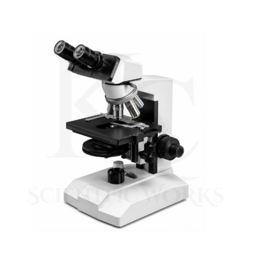
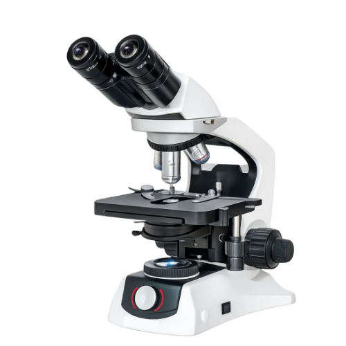
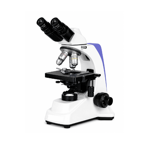
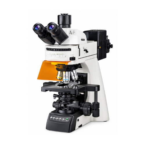
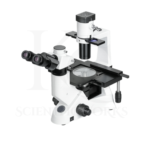
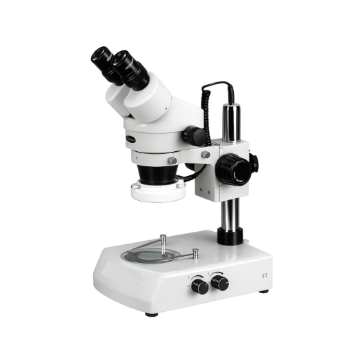
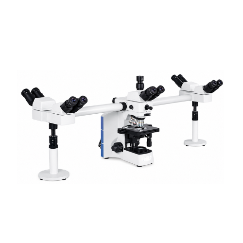
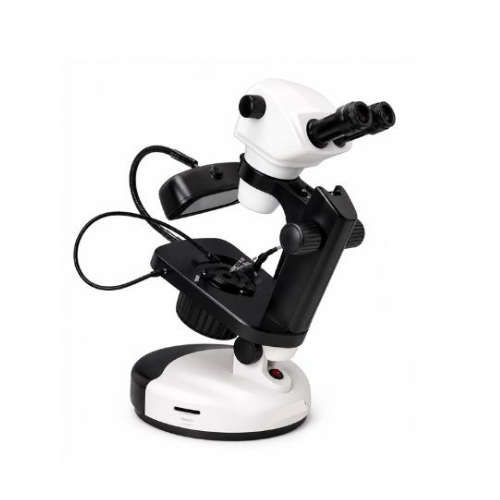
The Phase Contrast Microscope KCX-200 is intended for examining transparent and low-contrast biological specimens.
Many biological specimens contain structures that are difficult to distinguish using conventional bright-field illumination because they absorb very little light.
Phase contrast microscopy uses differences in optical path length to create visible image contrast.
This technique can help researchers observe suitable unstained cells, microorganisms and other transparent biological specimens.
The KCX-200 reference specifies Plan DIN Achromatic objectives, a sliding binocular viewing head, a double-layer mechanical stage, an Abbe condenser and precision coaxial focusing.
The microscope offers multiple illumination configurations, including halogen, incandescent, fluorescent-lamp and LED options.
Its optical magnification range depends on the installed eyepieces and objective lenses.
The KCX-200 is intended for biological education, cell observation, microbiology, tissue culture research and appropriate laboratory microscopy.
Important: Functional phase contrast observation requires compatible phase objectives and matched phase annuli or a suitable phase contrast condenser arrangement. These components are not detailed in the supplied reference and must be confirmed for the final KCX-200 configuration.
Applications
1. Cell Biology
The KCX-200 Phase Contrast Microscope can support examination of suitable transparent cell specimens.
2. Microbiology
A correctly configured phase contrast system can assist in observation of appropriate microorganisms.
3. Biotechnology Laboratories
The microscope is suitable for relevant biological microscopy applications in biotechnology research.
4. Tissue Culture Laboratories
Phase contrast microscopy can support examination of suitable cell cultures and unstained specimens.
5. Biological Research
The KCX-200 can be used for observation of compatible biological samples.
6. Pharmaceutical Research
The microscope can support appropriate specimen observation in pharmaceutical research environments.
7. Medical Research
Research laboratories can use phase contrast microscopy for suitable biological investigations.
8. Clinical Laboratory Research
The microscope can support research and educational microscopy involving prepared biological samples.
9. Universities
The KCX-200 is suitable for advanced biological microscopy education.
10. Medical Colleges
The microscope can support teaching of phase contrast microscopy principles and specimen observation.
11. Research Institutions
The KCX-200 can be used for suitable microscopy research applications.
12. Life Science Laboratories
The microscope supports observation of transparent and low-contrast biological specimens.
13. Educational Laboratories
Students can learn optical microscopy, specimen preparation and focusing techniques.
14. Environmental Microbiology
Phase contrast microscopy can support examination of suitable environmental microorganism samples.
15. Water Microbiology
The microscope can be used for observation of appropriate microorganisms in prepared water samples.
16. Pharmaceutical Laboratories
The KCX-200 can support suitable microscopy-based laboratory research and training.
17. Quality-Control Laboratories
The microscope can be used for applicable optical inspection and specimen observation.
18. Unstained Cell Observation
Phase contrast microscopy supports visualization of suitable cells without routine staining.
19. Microorganism Observation
The microscope can assist in examining compatible transparent microorganisms.
20. Scientific Microscopy Training
The KCX-200 supports practical training in phase contrast and transmitted-light microscopy.
Typical Specimens and Observations
| Specimen | Observation Purpose |
|---|
| Living Cells | Observation of suitable cell structures |
| Unstained Biological Specimens | Visualization of low-contrast features |
| Cell Cultures | Observation of cultured cells |
| Microorganisms | Examination of compatible microscopic organisms |
| Bacteria | Observation of suitable prepared bacterial specimens |
| Yeast | Examination of yeast cell structures |
| Protozoa | Observation of transparent microorganisms |
| Tissue Culture Samples | Observation of suitable cultured specimens |
| Thin Biological Specimens | Microscopic structural examination |
| Transparent Cellular Structures | Phase contrast visualization |
Phase Contrast Observation System
Phase contrast microscopy is an optical technique used to convert differences in the phase of transmitted light into visible differences in image intensity.
It is particularly useful for transparent specimens that show limited contrast under ordinary bright-field illumination.
A complete phase contrast system normally includes compatible phase contrast objectives and corresponding condenser annuli.
The optical configuration must be properly matched and aligned for effective phase contrast observation.
Phase Contrast System Details
| Parameter | Specification |
|---|
| Microscope Category | Phase Contrast |
| Optical Objectives | Plan DIN Achromatic |
| Listed Objectives | 4X, 10X, 40X and 100X |
| Condenser | Abbe, N.A. 1.25 |
| Phase Objectives | Not Individually Specified |
| Phase Annuli | Not Specified |
| Phase Centering Mechanism | Not Specified |
| Phase Telescope | Not Specified |
| Bright-Field Observation | Supported by Listed Optical Configuration |
| Phase Contrast Operation | Requires Confirmed Compatible Phase Components |
Optical System and Objectives
| Objective | Configuration | Magnification with WF10X | Magnification with WF16X |
|---|
| 4X | Plan DIN Achromatic | 40X | 64X |
| 10X | Plan DIN Achromatic | 100X | 160X |
| 40X | Plan DIN Achromatic, Spring-Loaded | 400X | 640X |
| 100X | Plan DIN Achromatic, Spring-Loaded, Oil | 1000X | 1600X |
The listed objective specifications describe the reference optical configuration. Their phase contrast compatibility must be verified separately.
Magnification System
Total optical magnification is calculated using:
Total Magnification = Eyepiece Magnification × Objective Magnification
Standard WF10X Configuration
| Eyepiece | Objective | Total Magnification |
|---|
| WF10X | 4X | 40X |
| WF10X | 10X | 100X |
| WF10X | 40X | 400X |
| WF10X | 100X | 1000X |
Optional WF16X Configuration
| Eyepiece | Objective | Total Magnification |
|---|
| WF16X | 4X | 64X |
| WF16X | 10X | 160X |
| WF16X | 40X | 640X |
| WF16X | 100X | 1600X |
Maximum stated nominal magnification: 1600X with the optional WF16X eyepiece and 100X objective.
Mechanical Stage
The KCX-200 features a double-layer mechanical stage for controlled positioning of microscope slides.
Stage Specifications
| Parameter | Specification |
|---|
| Stage Type | Double-Layer Mechanical |
| Stage Dimensions | 140 × 155 mm |
| X-Y Movement | 70 × 50 mm |
| Primary Function | Specimen Positioning |
| Suitable Specimens | Compatible Prepared Microscope Slides |
Condenser and Diaphragm System
The microscope includes an Abbe condenser with numerical aperture 1.25.
The condenser supports transmitted-light illumination by directing light toward the specimen.
The iris diaphragm allows adjustment of the illumination aperture.
Condenser Specifications
| Parameter | Specification |
|---|
| Condenser Type | Abbe |
| Numerical Aperture | 1.25 |
| Iris Diaphragm | Ø2–Ø30 mm |
| Optical Filter | Ø32 mm |
| Phase Annulus Arrangement | Not Specified |
Focusing System
The KCX-200 Phase Contrast Microscope incorporates coaxial coarse and fine focusing controls.
The coarse focusing mechanism supports initial specimen focusing.
The fine focusing mechanism allows smaller adjustments for image clarity.
Focusing Specifications
| Parameter | Specification |
|---|
| Focusing Type | Coaxial Coarse and Fine |
| Total Focusing Travel | 25 mm |
| Fine Focusing Precision | 0.002 mm |
| Fine Focusing Precision in Micrometers | 2 µm |
Illumination Configurations
The reference specifies several alternative illumination options.
| Illumination Type | Rating |
|---|
| Halogen | 6V / 20W |
| Incandescent | 20W |
| Fluorescent Lamp | 5W |
| LED | 1W |
| Optional Rechargeable LED | Available as Referenced |
These are alternative configurations, not necessarily lamps supplied together with one microscope.
Working Principle
The Phase Contrast Microscope KCX-200 is intended to operate using the principle of phase contrast microscopy.
Transparent biological specimens can alter the phase of transmitted light without absorbing much of it.
Under ordinary bright-field illumination, these phase differences may be difficult to observe.
A properly configured phase contrast microscope uses a condenser annulus and a corresponding phase plate in the objective.
The illumination system produces a hollow cone of light directed toward the specimen.
Light passing through the specimen is affected by differences in refractive index and thickness.
The phase contrast optical system causes appropriate interference between direct and diffracted light.
This converts optical phase differences into visible intensity differences.
As a result, suitable transparent structures become easier to distinguish.
The microscope's mechanical stage allows controlled positioning of the specimen.
The coaxial focusing system is used to obtain a clear image.
Basic Phase Contrast Observation Process
Specimen Preparation: Prepare a suitable transparent or low-contrast specimen.
Slide Positioning: Place the specimen on the mechanical stage.
Illumination: Switch on the selected transmitted-light illumination system.
Objective Selection: Select the required compatible objective.
Initial Focusing: Locate the specimen using coarse and fine focusing controls.
Phase Component Selection: Select the matching phase condenser annulus where fitted.
Phase Alignment: Align the phase optical system using the appropriate supplied method.
Observation: Examine the specimen through the binocular eyepieces.
Stage Movement: Move the specimen using the mechanical stage controls.
Magnification Adjustment: Change objectives as required, using the corresponding compatible phase components.
Educational and Research Benefits
The KCX-200 Phase Contrast Microscope supports training and research involving optical microscopy.
It helps users study:
Phase contrast microscopy principles
Transparent specimen observation
Unstained biological specimens
Cell biology
Microbiology
Tissue culture observation
Optical magnification
Objective selection
Condenser adjustment
Coaxial focusing
Mechanical stage operation
Biological specimen preparation
Bright-field and phase contrast differences
Laboratory microscopy techniques
Microscope Compatibility and Usage
The KCX-200 Phase Contrast Microscope is intended for appropriate biological and laboratory microscopy applications.
It can be used in research institutions, universities, biotechnology laboratories, microbiology departments and educational facilities.
The microscope should be placed on a stable laboratory surface.
Specimens should be prepared using appropriate laboratory procedures.
The illumination configuration should be selected according to the supplied model.
Observation should begin with a lower-power objective.
Coarse and fine focusing controls should be adjusted carefully.
For phase contrast microscopy, the installed phase objectives and condenser annuli must be correctly matched.
The optional WF16X eyepieces can be used where compatible with the supplied optical configuration.
The 100X oil immersion objective should be used with suitable immersion oil.
After use, the optical components should be cleaned with appropriate optical cleaning materials.
The microscope should be stored in a clean, dry environment.