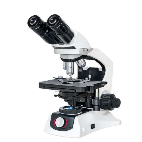
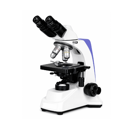
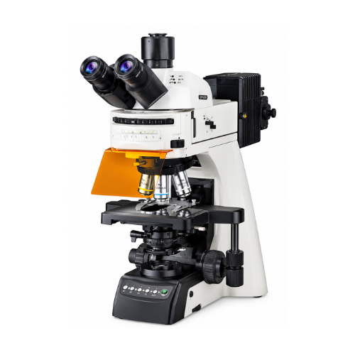
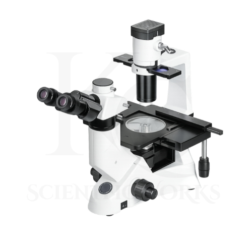
The Research Trinocular Microscope Max Series KCX-130 is designed for microscopic examination of biological specimens, microorganisms, prepared tissue sections, blood smears and suitable laboratory samples.
Its infinity-corrected optical system supports detailed specimen imaging.
The microscope incorporates plan achromatic objectives with magnifications of 4X, 10X, 40X and 100X oil immersion.
These objectives, combined with WF10X eyepieces, provide nominal magnifications from 40X to 1000X.
The trinocular observation head allows users to examine specimens directly and connect compatible camera equipment when required.
The rackless mechanical stage supports controlled specimen movement.
The coaxial coarse and fine focusing system allows users to adjust image sharpness.
The 5W LED Köhler illumination system provides adjustable transmitted-light illumination.
The KCX-130 is intended for biological research laboratories, pathology departments, microbiology laboratories, medical colleges, universities, hospitals and pharmaceutical research facilities.
Applications
1. Research Laboratories
The KCX-130 Research Trinocular Microscope Max Series supports detailed examination of prepared biological specimens during scientific research.
2. Medical Colleges
The microscope supports practical teaching of biological and medical microscopy.
3. Universities
The KCX-130 is suitable for laboratory education and biological research activities.
4. Pathology Laboratories
The microscope can be used for observation of prepared pathology slides.
5. Histopathology Laboratories
The KCX-130 supports examination of prepared histological tissue sections.
6. Microbiology Laboratories
The microscope can be used to observe appropriately prepared microbial specimens.
7. Pharmaceutical Industries
The KCX-130 supports suitable microscopic examination in pharmaceutical laboratory environments.
8. Biotechnology Research
The microscope can assist with biological specimen observation in biotechnology projects.
9. Clinical Diagnostic Laboratories
The microscope supports specimen examination in appropriate clinical laboratory workflows.
10. Food Testing Laboratories
The KCX-130 can support suitable microscopy-based food testing activities.
11. Environmental Testing Laboratories
The microscope can be used for observation of appropriate environmental samples and prepared slides.
12. Veterinary Laboratories
The KCX-130 supports microscopic examination of prepared veterinary specimens.
13. Quality-Control Laboratories
The microscope can assist with appropriate microscopy-based quality-control activities.
14. Hematology Laboratories
The KCX-130 supports examination of properly prepared blood smears.
15. Cell Biology Laboratories
The microscope can be used for observation of suitable cellular specimens.
16. Botany Laboratories
The KCX-130 supports examination of prepared plant tissues and cells.
17. Zoology Laboratories
The microscope can be used for observation of prepared animal tissues and specimens.
18. Digital Microscopy Documentation
The trinocular head supports compatible camera-based imaging for documentation.
19. Scientific Education and Training
The KCX-130 supports instruction in microscopy techniques and biological specimen examination.
20. Research and Development
The microscope is suitable for appropriate scientific projects involving microscopic specimen analysis.
Typical Specimens and Observations
| Specimen | Observation Purpose |
|---|
| Prepared Blood Smears | Blood cell morphology observation |
| Histology Slides | Tissue structure examination |
| Prepared Bacterial Smears | Microbial morphology observation |
| Plant Tissue Sections | Botanical structure examination |
| Animal Tissue Sections | Zoological and histological studies |
| Prepared Cell Slides | Cellular structure observation |
| Microbiology Teaching Slides | Educational microbial examination |
| Stained Tissue Sections | Histopathology studies |
| Veterinary Specimen Slides | Veterinary laboratory microscopy |
| Prepared Food Samples | Suitable microscopic examination |
| Environmental Specimen Slides | Microscopic observation |
| Biological Research Samples | Scientific specimen examination |
Working Principle
The Research Trinocular Microscope Max Series KCX-130 operates on the principle of transmitted-light compound microscopy.
Light from the built-in 5W LED illumination system is directed toward the prepared specimen.
The Köhler illumination arrangement allows the illumination to be adjusted for controlled specimen lighting.
Light passing through the specimen enters the selected objective lens.
The infinity-corrected optical system produces a magnified image through its compatible objective and tube-lens arrangement.
The image is viewed through the WF10X wide-field eyepieces.
The trinocular observation head provides an additional optical pathway for compatible digital imaging equipment.
Different objective magnifications can be selected using the quintuple revolving nosepiece.
The coaxial focusing controls allow adjustment of image sharpness.
The rackless mechanical stage supports controlled movement of the specimen during observation.
The 100X oil immersion objective is used with suitable immersion oil for appropriate high-magnification examination.
Basic Observation Procedure
Microscope Setup: Place the KCX-130 on a stable laboratory bench.
Power Connection: Connect the microscope to a suitable AC power source.
Specimen Preparation: Select an appropriate prepared microscope slide.
Slide Positioning: Position the slide on the mechanical stage.
Illumination: Switch on the 5W LED illumination system.
Köhler Adjustment: Adjust the illumination according to the microscope's operating procedure.
Objective Selection: Begin observation with the 4X objective.
Initial Focusing: Use coarse focusing to locate the specimen.
Fine Focusing: Adjust fine focus for a clear image.
Stage Movement: Use the mechanical stage controls to examine different specimen areas.
Higher Magnification: Select the 10X or 40X objective as required.
Oil Immersion: Use the 100X objective with appropriate immersion oil when necessary.
Digital Imaging: Connect compatible camera equipment if imaging is required.
Observation: Examine and document the relevant specimen characteristics.
Completion: Clean the oil immersion objective correctly after use and switch off the microscope.
Magnification System
The KCX-130 includes WF10X eyepieces and four specified plan achromatic objectives.
Total nominal magnification is calculated as:
Total Magnification = Eyepiece Magnification × Objective Magnification
| Objective | Eyepiece | Total Magnification |
|---|
| 4X | 10X | 40X |
| 10X | 10X | 100X |
| 40X | 10X | 400X |
| 100X Oil Immersion | 10X | 1000X |
Standard nominal magnification range: 40X–1000X.
The quintuple nosepiece provides five positions, but only four objective magnifications are specified.
Infinity-Corrected Optical System
The KCX-130 incorporates an infinity-corrected optical system with plan achromatic objectives.
Optical Specifications
| Parameter | Specification |
|---|
| Optical System | Infinity-Corrected |
| Objective Type | Plan Achromatic |
| Eyepieces | WF10X/22 mm |
| Objective Magnifications | 4X, 10X, 40X and 100X |
| Nosepiece | Quintuple Ball-Bearing |
| Nominal Magnification | 40X–1000X |
Infinity-corrected optics are designed to operate with a compatible tube-lens arrangement.
Plan achromatic objectives provide selected chromatic aberration correction and improved field flatness.
Trinocular Viewing System
The KCX-130 features a trinocular observation head for direct viewing and compatible digital imaging.
Viewing Specifications
| Parameter | Specification |
|---|
| Viewing Head | Trinocular |
| Inclination | 30° |
| Rotation | 360° |
| Eyepiece Magnification | 10X |
| Eyepiece Field Number | 22 mm |
| Camera Attachment | Trinocular Port |
| Interpupillary Adjustment | Not Specified |
| Diopter Adjustment | Not Specified |
Quintuple Ball-Bearing Nosepiece
The microscope incorporates a five-position revolving nosepiece.
Nosepiece Specifications
| Parameter | Specification |
|---|
| Nosepiece Type | Quintuple |
| Mechanism | Ball-Bearing |
| Objective Positions | 5 |
| Specified Objectives | 4X, 10X, 40X and 100X |
| Additional Objective | Not Specified |
| Primary Function | Objective Selection |
Rackless Mechanical Stage
The KCX-130 incorporates a rackless mechanical stage for controlled specimen positioning.
Stage Specifications
| Parameter | Specification |
|---|
| Stage Type | Rackless Mechanical |
| Specimen Movement | X-Y |
| Stage Dimensions | Not Specified |
| X-Axis Travel | Not Specified |
| Y-Axis Travel | Not Specified |
| Primary Function | Controlled Slide Positioning |
Coaxial Focusing System
The microscope features coaxial coarse and fine focusing controls.
Focusing Specifications
| Parameter | Specification |
|---|
| Focusing Type | Coaxial |
| Coarse Focusing | Provided |
| Fine Focusing | Provided |
| Fine Focus Division | Not Specified |
| Focusing Travel | Not Specified |
| Primary Function | Image Sharpness Adjustment |
5W LED Köhler Illumination System
The KCX-130 incorporates a 5W LED illumination system with adjustable intensity.
Illumination Specifications
| Parameter | Specification |
|---|
| Light Source | LED |
| LED Power | 5W |
| Illumination Method | Köhler |
| Brightness Control | Adjustable |
| Power Supply | AC Powered |
| Input Voltage | Not Specified |
| Primary Function | Transmitted-Light Illumination |
Benefits of Köhler Illumination
Köhler illumination is an optical illumination method used to achieve controlled and relatively even illumination across the specimen field.
When correctly adjusted, it helps reduce unwanted illumination artifacts and supports suitable contrast during biological microscopy.
Digital Imaging Compatibility
The KCX-130 features a trinocular observation port for compatible microscope cameras.
Imaging Specifications
| Parameter | Specification |
|---|
| Camera Port | Trinocular |
| Camera Attachment | Compatible Adapter Required |
| Camera Resolution | Not Specified |
| Camera Interface | Not Specified |
| Imaging Software | Not Specified |
| Camera Included | Not Confirmed |
| Camera Adapter Included | Not Confirmed |
Digital Microscopy Applications
With a suitable camera and adapter, the microscope can support:
Digital specimen photography
Laboratory image documentation
Live microscope viewing on a compatible display
Educational demonstrations
Research image archiving
Scientific presentation preparation
Body Construction and Electrical System
The KCX-130 features a heavy-duty metal body with a corrosion-resistant powder-coated finish.
Construction Specifications
| Parameter | Specification |
|---|
| Body Material | Metal |
| Construction | Heavy-Duty |
| Finish | Powder Coated |
| Corrosion Protection | Corrosion-Resistant Finish |
| Illumination Power | AC |
| Built-In Lighting | 5W LED |
Educational and Research Benefits
The KCX-130 Research Trinocular Microscope Max Series supports:
Biological specimen examination
Microbiology laboratory observation
Histology studies
Prepared blood smear examination
Cellular morphology studies
Tissue structure observation
Plant tissue examination
Animal tissue microscopy
Medical laboratory education
Veterinary laboratory microscopy
Biotechnology research
Pharmaceutical laboratory applications
Scientific laboratory training
Digital microscopy documentation
Oil immersion microscopy
Infinity optical system training
Mechanical stage operation
Precision focusing
Köhler illumination adjustment
Advanced research microscopy
Microscope Compatibility and Usage
The KCX-130 Research Trinocular Microscope Max Series is intended for biological, educational, clinical and research laboratory applications.
The microscope should be placed on a stable laboratory bench.
Prepared slides should be positioned correctly on the mechanical stage.
The LED illumination system should be adjusted according to the specimen and selected objective.
Observation should begin with the 4X objective.
The coaxial focusing controls should be used carefully.
The mechanical stage allows controlled movement of the specimen.
Higher-magnification objectives can be selected when required.
The 100X oil immersion objective should be used only with suitable immersion oil.
The objective should be cleaned correctly after oil immersion use.
The trinocular port can be used for compatible digital camera attachment when required.
After observation, the microscope should be switched off and stored in a clean, dry environment.