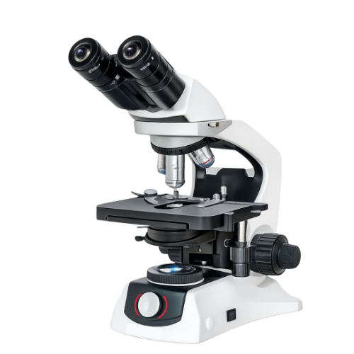
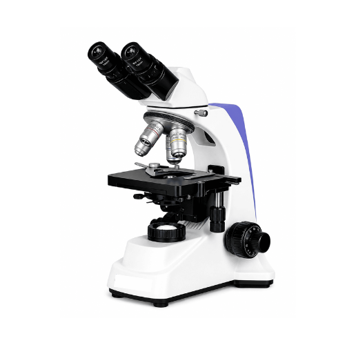
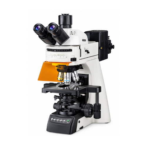
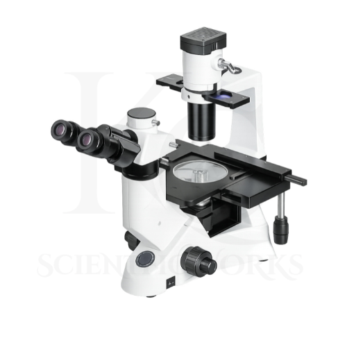
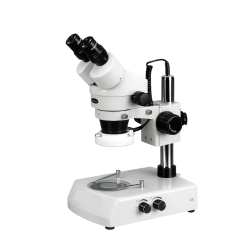
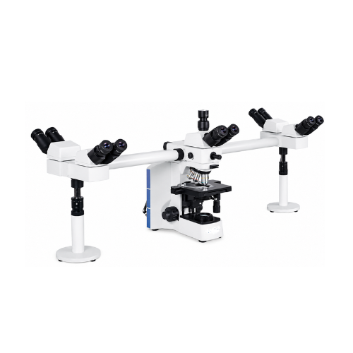
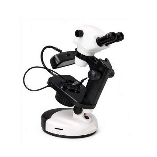
The Tissue Culture Microscope KCX-168 is intended for observation and imaging of cultured cells, biological tissues and suitable laboratory specimens.
Unlike conventional upright microscopes, the inverted optical system positions the objectives below the specimen stage.
This arrangement is useful for examining cells located near the bottom of compatible culture vessels.
Brightfield microscopy provides transmitted-light observation of suitable specimens.
Phase contrast microscopy improves the visibility of low-contrast structures without requiring fluorescence labeling.
Fluorescence microscopy allows observation of compatible fluorescent labels using suitable excitation and emission filters.
The trinocular observation head supports visual examination and digital imaging.
The mechanical stage allows specimen positioning.
The scientific CMOS camera supports digital image acquisition according to the camera's verified performance.
The imaging software provides tools for fluorescence analysis, measurement, annotation and cell counting.
The KCX-168 is intended for cell culture research, biotechnology, pharmaceutical research, academic laboratories and suitable biological imaging workflows.
Applications
1. Tissue Culture Laboratories
The KCX-168 Tissue Culture Microscope supports observation of suitable cultured cell specimens.
2. Live Cell Imaging
The microscope can be used for imaging living cells under appropriate laboratory conditions.
3. Cell Biology Research
The KCX-168 supports examination of cellular morphology and biological structures.
4. Biotechnology Laboratories
The microscope supports suitable biotechnology research and imaging activities.
5. Pharmaceutical Research
The KCX-168 can be used for cell-based research observation.
6. Fluorescence Microscopy
The microscope supports imaging of appropriately fluorescently labeled specimens.
7. Phase Contrast Microscopy
The KCX-168 supports observation of suitable low-contrast cell specimens.
8. Brightfield Microscopy
The microscope can be used for conventional transmitted-light specimen observation.
9. Cell Culture Monitoring
The KCX-168 supports visual monitoring of suitable cell cultures.
10. Cell Morphology Studies
The microscope can be used to examine cellular shape and structural changes.
11. Fluorescence Channel Imaging
The KCX-168 supports imaging using compatible DAPI, FITC and TRITC filter sets.
12. Time-Lapse Research
The reference software supports repeated image capture for suitable time-lapse experiments.
13. Cell Counting
The microscope's specified software includes automatic and manual cell counting tools.
14. Tissue Research
The KCX-168 supports observation of suitable biological tissue specimens.
15. Academic Research Laboratories
The microscope can be used for advanced biological microscopy education and research.
16. Molecular Biology Research
The KCX-168 supports observation of suitably labeled biological specimens.
17. Immunofluorescence Studies
The microscope can be used with appropriate fluorescence-labeled samples.
18. Biomedical Research
The KCX-168 supports suitable non-diagnostic biomedical imaging activities.
19. Cell-Based Assays
The microscope can be used for visual analysis in suitable cell-based research workflows.
20. Scientific Image Documentation
The camera and imaging software support digital recording and analysis of microscope images.
Typical Specimens and Observations
| Specimen | Observation Purpose |
|---|
| Adherent Cell Cultures | Cell morphology observation |
| Cultured Mammalian Cells | Live cell imaging |
| Cell Culture Dishes | Inverted brightfield observation |
| Cell Culture Flasks | Cell growth examination |
| Fluorescently Labeled Cells | Fluorescence imaging |
| DAPI-Labeled Specimens | Compatible fluorescence observation |
| FITC-Labeled Specimens | Compatible fluorescence observation |
| TRITC-Labeled Specimens | Compatible fluorescence observation |
| Transparent Biological Samples | Phase contrast observation |
| Suitable Tissue Samples | Biological structure examination |
| Cell-Based Research Samples | Imaging and analysis |
| Prepared Laboratory Slides | Compatible optical observation |
Working Principle
The Tissue Culture Microscope KCX-168 operates on the principle of inverted optical microscopy with brightfield, phase contrast and fluorescence observation.
The objective lenses are positioned below the specimen stage.
Suitable specimens are placed in compatible culture vessels or specimen holders.
For brightfield observation, transmitted light passes through the specimen and is collected by the objective.
For phase contrast observation, compatible phase optics convert phase differences in the specimen into observable intensity variations.
For fluorescence observation, excitation light passes through the appropriate optical filter arrangement and reaches the labeled specimen.
Fluorescent molecules emit light at wavelengths determined by their optical properties.
The fluorescence filter system helps separate the desired emission from excitation light.
The objective collects the specimen image, which is directed toward the binocular viewing system or camera path.
The scientific CMOS camera converts the optical image into digital image data.
The software processes the images for visualization, measurement and analysis.
Basic Observation Procedure
Microscope Setup: Place the KCX-168 on a stable laboratory surface.
System Inspection: Check the microscope, stage, objectives and imaging connections.
Illumination Setup: Activate the appropriate illumination system.
Specimen Selection: Choose a suitable prepared specimen or culture vessel.
Specimen Placement: Position the specimen using the appropriate holder.
Observation Mode: Select brightfield, phase contrast or fluorescence.
Objective Selection: Begin with an appropriate lower-power objective.
Initial Focusing: Bring the specimen into focus using the microscope's focusing system.
Eyepiece Adjustment: Adjust the viewing system for comfortable observation.
Stage Positioning: Move the mechanical stage to locate the desired specimen area.
Image Optimization: Adjust illumination and contrast according to the selected mode.
Fluorescence Filter Selection: Select the appropriate filter set for fluorescent specimens.
Camera Activation: Connect the scientific CMOS camera to the imaging computer.
Software Setup: Open the compatible imaging software.
Image Capture: Acquire images using suitable exposure settings.
Image Analysis: Apply the required measurement or fluorescence analysis tools.
Data Recording: Save the acquired images and associated observations.
Completion: Turn off illumination and secure the microscope after use.
Fluorescence illumination and live-cell handling should follow the laboratory's approved safety and specimen-care procedures.
Magnification System
The KCX-168 reference specifies 10X eyepieces and Plan Achromat / Plan Fluor long-working-distance objectives.
However, the individual objective magnifications are not provided.
Total Visual Magnification = Eyepiece Magnification × Objective Magnification
| Optical Component | Specification |
|---|
| Eyepiece Magnification | 10X |
| Objective Type | Plan Achromat / Plan Fluor LWD |
| Standard Objective Magnifications | Not Specified |
| Nosepiece | Encoded Quadruple |
| Optional Objective | Up to 100X Oil Immersion |
| Standard Total Magnification Range | Not Confirmed |
For example, a compatible 20X objective combined with a 10X eyepiece would produce 200X nominal visual magnification.
This is a mathematical example, not confirmation that a 20X objective is supplied with KCX-168.
Infinity-Corrected Optical System
The microscope incorporates an infinity-corrected optical design.
Optical Specifications
| Parameter | Specification |
|---|
| Optical System | Infinity Corrected |
| Viewing Head | Trinocular |
| Eyepieces | 10X / FN 23 mm |
| Objectives | Plan Achromat and Plan Fluor LWD |
| Nosepiece | Encoded Quadruple |
| Observation Modes | Brightfield, Phase Contrast and Fluorescence |
| DIC | Optional |
| Emboss Contrast | Optional |
Trinocular Viewing System
The KCX-168 features a trinocular observation head.
Viewing Specifications
| Parameter | Specification |
|---|
| Observation Head | Trinocular |
| Light Distribution | 100:0 / 0:100 |
| Eyepieces | 10X |
| Field Number | 23 mm |
| Diopter Adjustment | Provided |
| Anti-Fungus Treatment | Specified |
| Interpupillary Distance | Not Specified |
| Head Inclination | Not Specified |
Long-Working-Distance Objective System
The microscope reference includes Plan Achromat and Plan Fluor LWD objectives.
Objective Specifications
| Parameter | Specification |
|---|
| Objective Type | Plan Achromat / Plan Fluor |
| Working Distance Category | Long Working Distance |
| Optical Correction | Infinity |
| Standard Magnifications | Not Specified |
| Phase Contrast Compatibility | Referenced |
| Fluorescence Compatibility | Referenced |
| Optional High Magnification | Up to 100X Oil Immersion |
ELWD Condenser and Phase Contrast
The KCX-168 reference specifies a focusable ELWD condenser with a centerable phase slider.
Condenser Specifications
| Parameter | Specification |
|---|
| Condenser Type | Extra Long Working Distance |
| Focusing | Provided |
| Numerical Aperture | 1.25 (Reference) |
| Phase Slider | Centerable |
| Phase Contrast Observation | Supported |
Verification note: An ELWD condenser with N.A. 1.25 is an unusual combination. The numerical aperture and working-distance specification should be checked against the final condenser model.
Mechanical Stage and Specimen Holder
The microscope incorporates an attachable mechanical stage.
Stage Specifications
| Parameter | Specification |
|---|
| Stage Type | Mechanical |
| Attachment | Attachable |
| Specimen Holder | Universal |
| Culture Dish Compatibility | Referenced |
| Culture Flask Compatibility | Referenced |
| Slide Compatibility | Referenced |
| X-Y Travel | Not Specified |
| Stage Dimensions | Not Specified |
| Motorized Stage | Not Specified |
LED Transmitted Illumination
The KCX-168 reference specifies a cool white LED illumination system.
Illumination Specifications
| Parameter | Specification |
|---|
| Illumination Type | Transmitted LED |
| Light Colour | Cool White |
| LED Power | 20–30 W |
| Claimed LED Lifespan | Over 60,000 Hours |
| Brightness Uniformity | Referenced |
| Brightness Adjustment Range | Not Specified |
| Electrical Supply | Not Specified |
Epi-Fluorescence Imaging System
The KCX-168 incorporates a reference epi-fluorescence attachment with a rotating filter turret.
Fluorescence Specifications
| Parameter | Specification |
|---|
| Fluorescence Type | Epi-Fluorescence |
| Filter Turret | Rotating |
| Number of Filter Cubes | Up to 3 |
| Filter Set 1 | DAPI |
| Filter Set 2 | FITC |
| Filter Set 3 | TRITC |
| Fluorescence Illumination | LED |
| Claimed Fluorescence LED Lifespan | Over 10,000 Hours |
| Synchronized LED / Filter Switching | Referenced |
| Automatic Intensity Recognition | Referenced |
DAPI Channel
The DAPI filter set is intended for observation of specimens labeled with compatible blue-emitting fluorescent markers.
FITC Channel
The FITC filter set supports imaging of specimens labeled with compatible green-emitting fluorophores.
TRITC Channel
The TRITC filter set supports observation of compatible orange-red fluorescent labels.
Actual excitation and emission wavelengths depend on the installed filter cubes and should be confirmed separately.
Scientific CMOS Camera
The KCX-168 reference configuration includes a 5.9 MP scientific CMOS camera.
Camera Specifications
| Parameter | Specification |
|---|
| Camera Type | Scientific CMOS |
| Resolution | 5.9 MP |
| Interface | USB 3.0 |
| Quantum Efficiency | 60% (Reference) |
| Live Frame Rate | 10–15 fps (Reference) |
| Long Exposure | Supported |
| Sensor Size | Not Specified |
| Pixel Size | Not Specified |
| Cooling | Not Specified |
| Exposure Range | Not Specified |
| Camera Model | Not Specified |
The 10–15 fps value is not associated with a specified resolution or acquisition mode in the reference. Model-specific camera documentation should establish the verified frame rate.
Advanced Imaging and Fluorescence Analysis Software
The reference software includes digital imaging and fluorescence analysis functions.
Software Features
| Function | Reference Capability |
|---|
| Live Image Display | Supported |
| Image Capture | Supported |
| Fluorescence Channel Mixing | Supported |
| Fluorescence Channel Unmixing | Referenced |
| Time-Lapse Imaging | Supported |
| Manual Cell Counting | Supported |
| Automatic Cell Counting | Supported |
| Length Measurement | Supported |
| Area Measurement | Supported |
| Perimeter Measurement | Supported |
| Intensity Measurement | Supported |
| Image Enhancement | Supported |
| Image Overlay | Supported |
| Annotation | Supported |
| Shading Correction | Supported |
These functions should be verified against the final supplied software version and license.
Computer System Configuration
The supplied reference includes the following computer configuration.
| Parameter | Specification |
|---|
| Processor | Intel Core i5, 13th Generation |
| RAM | 16 GB |
| Storage | 2 TB HDD |
| Operating System | Windows 10 |
| Camera Connectivity | USB 3.0 |
| Imaging Software | Advanced Imaging and Fluorescence Analysis |
Computer inclusion, exact hardware model and operating-system compatibility should be confirmed in the final supply scope.
Educational and Research Benefits
The KCX-168 Tissue Culture Microscope supports:
Tissue culture observation
Live cell imaging
Cell morphology examination
Brightfield microscopy
Phase contrast microscopy
Fluorescence microscopy
DAPI fluorescence observation
FITC fluorescence observation
TRITC fluorescence observation
Digital image acquisition
Scientific image documentation
Cell counting
Fluorescence intensity analysis
Image measurement
Time-lapse imaging
Cell-based research
Biotechnology studies
Pharmaceutical research
Academic microscopy training
Biological image analysis
Microscope Compatibility and Usage
The KCX-168 Tissue Culture Microscope is intended for research laboratories, biotechnology facilities, cell biology laboratories and academic institutions.
Suitable specimens should be placed in compatible culture vessels.
The universal specimen holder should be selected according to the vessel dimensions.
The appropriate objective should be selected for the required observation mode.
Brightfield illumination can be used for conventional transmitted-light observation.
Phase contrast can be used for suitable low-contrast specimens.
Fluorescence observation requires appropriate fluorescent labeling and compatible filter sets.
The scientific CMOS camera can be used for digital image capture according to its verified specifications.
The imaging software can be used for measurement, annotation and fluorescence image processing.
For live-cell experiments, environmental control requirements such as temperature, humidity and gas composition must be addressed separately. An environmental incubation chamber is not specified in the supplied reference.