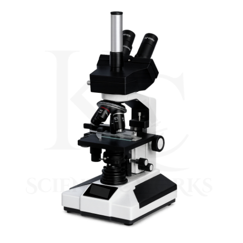
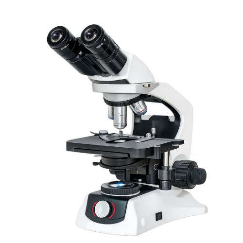
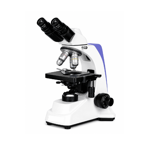
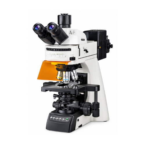
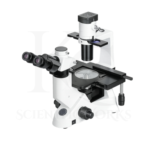
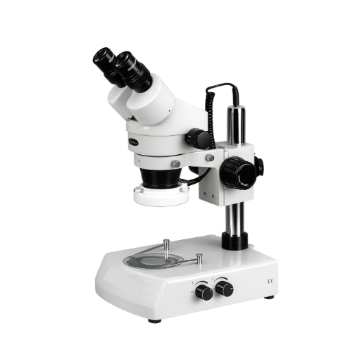
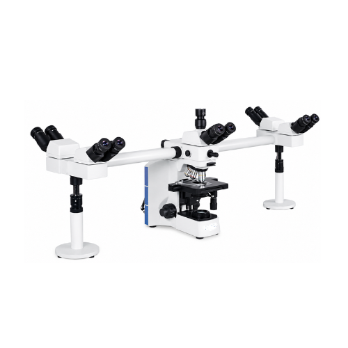
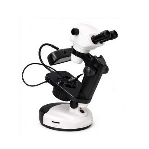
The Trinocular Microscope KCX-140 is designed to provide magnified optical observation and digital documentation of microscopic specimens.
The microscope uses semi-plan achromatic objectives to produce magnified images of suitable prepared specimens.
The WF10X and WF15X wide-field eyepieces provide visual observation through the trinocular viewing head.
The 45° inclined observation head supports routine laboratory viewing.
The LED illumination system provides light for transmitted-light specimen observation.
The coarse focusing mechanism allows initial focusing.
The fine focusing mechanism supports more precise image adjustment.
The integrated 1.3 MP CMOS camera provides a digital imaging path.
The USB interface allows connection to compatible computer equipment.
The microscope is intended for biological research, microbiology education, pathology teaching, laboratory demonstrations and scientific image documentation.
Applications
1. Biological Research Laboratories
The KCX-140 Trinocular Microscope supports observation of suitable biological specimens.
2. Microbiology Laboratories
The microscope can be used for observation of properly prepared microbiological slides.
3. Pathology Teaching Laboratories
The KCX-140 supports educational examination of prepared pathology specimens.
4. Histology Laboratories
The microscope can be used for observation of suitable tissue sections.
5. School Biology Laboratories
The KCX-140 supports advanced school-level microscopy education.
6. College Science Laboratories
The microscope can be used for practical biology exercises.
7. University Research Laboratories
The KCX-140 supports suitable academic microscopy activities.
8. Medical Education
The microscope can be used for non-diagnostic examination of prepared teaching slides.
9. Pharmaceutical Research Laboratories
The KCX-140 supports suitable microscopic observation of laboratory samples.
10. Biotechnology Laboratories
The microscope can be used for appropriate biological specimen examination.
11. Plant Biology Studies
The KCX-140 supports observation of plant tissue sections.
12. Animal Tissue Observation
The microscope can be used to examine prepared animal tissue specimens.
13. Microorganism Observation
The KCX-140 supports observation of suitably prepared microbial samples.
14. Blood Smear Teaching
The microscope can be used for educational observation of prepared blood smear slides.
15. Scientific Image Documentation
The camera supports digital capture of microscope images.
16. Laboratory Demonstrations
The KCX-140 can be used to demonstrate compound microscope operation.
17. Digital Microscopy Education
The microscope supports instruction in camera-based image acquisition.
18. Biological Specimen Comparison
The KCX-140 can be used to compare suitable prepared specimens.
19. Research Image Recording
The microscope supports digital recording for appropriate research activities.
20. Scientific Training Programs
The KCX-140 supports training in optical microscopy and digital image documentation.
Typical Specimens and Observations
| Specimen | Observation Purpose |
|---|
| Plant Tissue Slides | Cellular structure observation |
| Animal Tissue Sections | Histology education |
| Prepared Bacterial Slides | Microbiology teaching |
| Blood Smear Slides | Non-diagnostic hematology education |
| Onion Epidermis | Plant cell structure |
| Leaf Sections | Botanical microscopy |
| Prepared Microorganism Slides | Microbial morphology |
| Histology Teaching Slides | Tissue structure examination |
| Biological Research Slides | General specimen observation |
| Prepared Laboratory Samples | Scientific image documentation |
Working Principle
The Trinocular Microscope KCX-140 operates using transmitted-light compound microscopy and digital imaging.
Light from the LED illumination system passes through a suitable prepared specimen.
The objective lens collects light from the specimen and produces a magnified intermediate image.
The eyepiece magnifies this image for visual observation.
The trinocular viewing head provides an additional optical path for camera-based imaging.
The CMOS camera converts the optical image into digital image data.
The USB interface transfers supported image data to compatible computer equipment.
The coarse focusing mechanism adjusts the microscope for initial specimen observation.
The fine focusing mechanism supports precise adjustment of image sharpness.
The resulting optical and digital images can be used for suitable laboratory observation, teaching and scientific documentation.
Basic Observation Procedure
Microscope Setup: Place the KCX-140 on a stable laboratory surface.
Power Connection: Connect the microscope to a suitable 220V AC supply according to its electrical rating.
Optical Inspection: Check the eyepieces and objectives for cleanliness.
Illumination: Switch on the LED light source.
Specimen Selection: Choose a suitable prepared microscope slide.
Specimen Placement: Position the slide securely on the stage.
Eyepiece Selection: Select the appropriate WF10X or WF15X eyepieces.
Objective Selection: Begin with the lowest available objective magnification.
Initial Focusing: Use the coarse focusing control to locate the specimen.
Fine Focusing: Adjust the fine focus for a clear image.
Illumination Adjustment: Optimize specimen brightness using the available controls.
Magnification Change: Select a higher-power objective if required.
Focus Readjustment: Refine the image using fine focusing.
Camera Connection: Connect the built-in CMOS camera to a compatible computer through USB.
Software Setup: Open the supported camera imaging software.
Digital Observation: View the image using the available camera functions.
Image Capture: Capture suitable microscope images.
Documentation: Save images and record laboratory observations.
Completion: Remove the specimen and switch off the microscope.
Storage: Clean and store the microscope appropriately.
Magnification System
The KCX-140 reference specifies a total magnification range of 40X–1500X.
The microscope includes WF10X and WF15X eyepieces.
However, the individual objective magnifications are not specified.
Magnification Specifications
| Parameter | Specification |
|---|
| Stated Magnification Range | 40X–1500X |
| Eyepiece Option 1 | WF10X |
| Eyepiece Option 2 | WF15X |
| Objective Type | Semi-Plan Achromatic |
| Objective Magnifications | Not Specified |
| Optical Magnification Formula | Eyepiece × Objective |
For a conventional compound microscope:
Total Magnification = Eyepiece Magnification × Objective Magnification
Illustrative Magnification Combinations
The following table demonstrates common mathematical combinations. It does not establish which objectives are supplied with KCX-140.
| Example Objective | WF10X Eyepiece | WF15X Eyepiece |
|---|
| 4X | 40X | 60X |
| 10X | 100X | 150X |
| 40X | 400X | 600X |
| 100X | 1000X | 1500X |
Technical clarification: The stated 40X–1500X range is consistent with a possible 4X–100X objective arrangement, but the actual supplied objective configuration must be confirmed.
Trinocular Viewing System
The KCX-140 features a trinocular observation head.
Viewing Specifications
| Parameter | Specification |
|---|
| Observation Head | Trinocular |
| Viewing Angle | 45° Inclined |
| Eyepieces | WF10X and WF15X |
| Camera Path | Provided |
| Interpupillary Distance | Not Specified |
| Diopter Adjustment | Not Specified |
| Light Distribution Ratio | Not Specified |
| Head Rotation | Not Specified |
Semi-Plan Achromatic Objective System
The microscope incorporates semi-plan achromatic objectives.
Objective Specifications
| Parameter | Specification |
|---|
| Objective Type | Semi-Plan Achromatic |
| Optical Correction | Achromatic |
| Field Correction | Semi-Plan |
| Individual Magnifications | Not Specified |
| Numerical Aperture | Not Specified |
| Working Distance | Not Specified |
| Oil Immersion Objective | Not Confirmed |
Built-In 1.3 MP CMOS Camera
The KCX-140 includes a specified 1.3 MP CMOS camera.
Camera Specifications
| Parameter | Specification |
|---|
| Camera Type | CMOS |
| Camera Resolution | 1.3 MP |
| Camera Arrangement | Built-In |
| Interface | USB |
| Sensor Model | Not Specified |
| Pixel Size | Not Specified |
| Frame Rate | Not Specified |
| Exposure Control | Not Specified |
| White Balance | Not Specified |
| Image Measurement Software | Not Specified |
| Video Recording | Not Confirmed |
A 1.3 MP camera may provide a nominal image size of approximately 1280 × 1024 pixels, depending on the sensor. The exact output resolution must be verified against the installed camera model.
LED Illumination System
The KCX-140 features bright LED illumination.
Illumination Specifications
| Parameter | Specification |
|---|
| Light Source | LED |
| Observation Method | Transmitted Light |
| LED Power | Not Specified |
| Brightness Adjustment | Not Specified |
| Colour Temperature | Not Specified |
| LED Lifespan | Not Specified |
| Power Supply | 220V AC |
Precision Coarse and Fine Focusing
The microscope incorporates coarse and fine focusing.
Focusing Specifications
| Parameter | Specification |
|---|
| Coarse Focusing | Provided |
| Fine Focusing | Provided |
| Coaxial Controls | Not Specified |
| Fine Focus Resolution | Not Specified |
| Focusing Travel | Not Specified |
| Focus Stop | Not Specified |
Die-Cast Metal Construction
The KCX-140 features a die-cast metal body.
Construction Specifications
| Parameter | Specification |
|---|
| Body Material | Die-Cast Metal |
| Microscope Type | Trinocular Compound |
| Observation Head | 45° Inclined |
| Illumination | LED |
| Electrical Supply | 220V AC |
| Dimensions | Not Specified |
| Weight | Not Specified |
Digital Imaging and Computer Compatibility
The KCX-140 incorporates a USB-connected CMOS camera.
Digital Imaging Specifications
| Parameter | Specification |
|---|
| Camera | Built-In 1.3 MP CMOS |
| Interface | USB |
| Computer Connection | Supported with Compatible Hardware |
| Image Capture | Supported |
| Image Display | Subject to Camera Software |
| Image Measurement | Not Confirmed |
| Image Recording Format | Not Specified |
| Operating System Compatibility | Not Specified |
| Computer Included | Not Specified |
Educational and Laboratory Benefits
The KCX-140 Trinocular Microscope supports:
Biological specimen observation
Microbiology education
Pathology teaching
Histology examination
Scientific research
College biology practicals
University laboratory activities
Digital microscopy training
Camera-based image acquisition
Microscope image documentation
Plant cell observation
Animal tissue examination
Prepared microorganism observation
Optical magnification demonstrations
Objective lens training
Eyepiece magnification education
Coarse focusing practice
Fine focusing practice
LED microscopy demonstrations
Scientific image recording
Microscope Compatibility and Usage
The KCX-140 Trinocular Microscope is intended for research laboratories, educational institutions, microbiology departments and pathology teaching environments.
Suitable prepared slides should be used for specimen observation.
The microscope should be connected to an appropriate electrical supply.
The eyepieces and objectives should be selected according to the required magnification.
The LED illumination system should be adjusted to provide suitable specimen visibility.
The coarse focusing mechanism should be used for initial focusing.
The fine focusing mechanism should be used to refine the image.
The integrated CMOS camera can be connected to compatible computer equipment.
Digital images can be acquired using the appropriate supported software.
For diagnostic or clinical reporting, the microscope's intended use, performance and applicable regulatory compliance must be verified separately.